Rubtsov V. I., Krinichnaya E. P., Shul'ga Yu. M.
Institute of Problems of Chemical Physics, Russian Academy of Sciences, 142432 Chernogolovka, Moscow Region, Russia
Тел.: (095) 522-36-70, e-mail:shulga@icp.ac.ru (Shul'ga Yu. M.)
AUGER SPECTROSCOPY STUDY OF THE CARBON NANOMATERIAL ENRICHED BY NANOTUBES
Presence of chlorine atoms on the surface of powders, obtained by electric arc sputtering of the graphite-cobalt-nickel electrodes, after boiling of them in hydrochloric acid was found by Auger electron spectroscopy. Metallic atoms were observed in the analysis zone only after argon ion sputtering.
It is well known from microelectronics technology, that sputtering of silicon surface by solutions, containing the hydrochloric acid, leads to formation of stable surface chloride compounds. Special procedures to remove these compounds from the surface are necessary. So called "boiling" in concentrated hydrochloric acid is widely used to wash away the catalyst, inserted to sputtered electrode when the electric arc method is applied to obtain the carbon nan-otubes (see, for example, [1]). It is supposed that the hydrochloric acid does not interact with carbon material.
The aim of this paper is to study the surface of the carbon material obtained during the sputtering of graphite-cobalt-nickel electrode in electric arc, and then subjected to treatment which increases the content of single-wall carbon nanotubes. The treatment, one stage of which includes the boiling in concentrated hydrochloric acid, is described in experimental part. Auger electron spectroscopy was chosen due to high surface sensitivity to atomic relation in thin layer 10-30 A.
After boiling the colored liquid phase was separated from solid one, then it was poured out by fresh portion of the acid and was heated up to the same temperature. The cycle of the acid cleaning was repeated until the liquid phase stopped to stain.
Samples for Auger spectroscopy study were prepared by pressing of the powder to indium substrate. Auger spectra were registered with spectrometer PHI-551 equipped by double-pass cylindrical mirror analyzer. Base pressure in spectrometer chamber was less than 340"7 Pa.
For sputtering of sample surface by argon ions the test chamber was filled by argon with partial pressure 340"3 Pa. Auger spectra were registered immediately during sputtering. Peak-to-peak voltage amplitude was 3 V for kinetic energies up to 550 eV and 6 V for higher kinetic energies of electrons.
RESULTS AND DISCUSSION
EXPERIMENTAL
Method for obtaining of samples under study is analogous to one for fullerenes [2]. Graphite rod, which contained the catalyst, was used as anode. The catalyst was a mixture of cobalt and nickel powders with 3 cobalt atoms per 1 nickel one. Sputtered graphite rods with length of 150 mm and diameter of 8 mm had the channel along longitudinal axis. This channel had diameter of 3 mm and depth of 140 mm. Hollow part of the electrode was filled by catalyst. Content of catalyst in sputtered zone of the rod was approximately 15 mass. %.
Sputtering was carried out under helium pressure of 650 Torr. Discharge current was 100 A at voltage 2830 V. The distance between anode and cathode was equal to 2 mm during sputtering. Distance from electrodes to walls of chamber cooled by water was 70 mm.
The material obtained after sputtering was gathered from lateral walls of the chamber (so called parietal soot) and used for study. Enrichment procedure of the material by nanotubes was consisting in following: at first a sample was oxidized by dry air in muffle furnace with gradual slow increase of temperature up to 723 K to burn out amorphous carbon, and then one was boiled in concentrated hydrochloric acid at 358-368 K.
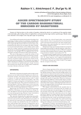
Survey Auger spectrum of the carbon material under study is presented in Fig. 1. It is seen that apart from carbon the chlorine atoms are observed on the sample surface. Oxygen turns to be absent in the surface layer. It is known [3], that opening of nanotube edges by sputtering in mixture of acids (H2SO4/HNO3) leads to formation of carboxyl (-COOH) groups on the surface of nanotubes. When the carbon material is partially oxidized, it must include oxygen-containing fragments. Absence of oxygen in the Auger analysis zone means that either oxygen-containing fragments on the surface are decomposed under electron beam, or its are in more deep layers of the carbon material.
The oxygen concentration in the analysis zone increases after top layer removing from the sample under study during argon ion sputtering. At the same time the intensity of the Auger line Cl LMM at 181 eV sharply diminishes. At that the cobalt lines arise in the Auger spectrum. Absence of nickel Auger lines may probably be explained by the fact, that the nickel concentration is 3 times less than the cobalt one following the preparation procedure. It is also possible that under sputtering the nickel forms opened particles, which are removed during the boiling in concentrated hydrochloric acid.
Evaluation of relative concentration [Co/C]at from peak intensity relation of the Auger lines Co LMV (780 eV) and C KVV (272 eV) gives the value 0.032. Analogous es-
E-mail: redactor@hydrogen.ru, http://www.hydrogen.ru
Rubtsov V. I., Krinichnaya E. P., Shul'ga Yu. M.
Auger Spectroscopy Study of the Carbon Nanomaterial Enriched by Nanotubes
18
a) b)
Fig. 1. Survey Auger spectra of powder, obtained by electric arc sputtering of graphite electrode with cobalt-nickel catalyst and enriched by single-wall nanotubes (see text for enrichment procedure) before (a) and after (b) argon ion sputtering. Boundary of modulation voltage change is marked in spectra by break
timation from the data on weighing the residue, which is incombustible in oxygen at 1670 K and ascribed as CoO, gives the value 0.055. If the metallic atoms were uniformly arranged in the carbon matrix or size of metal particles were less than mean free path of the Co LMV Auger electron, then the both evaluations of the relative concentration [Co/C] were close. Therefore we may suppose, that mean size of metal particles is more than mean free path X for electron with kinetic energy Ekin=780 eV in carbon matrix. The data on X may be taken from reference [4]. The universal dependence X = X(EUn) presents the value 4-5 monolayers for Ekin=800 eV. Calculation of this value following to Seach and Dench formula [5], which takes into account the influence of matrix on mean free path (X = 0.41 M15 E,05, where M - mean size of atom in matrix),
a kin' a '>
gives the value 1.1 nm Hence, mean size of metal particles in sample under study is more than 1.5-2.0 nm.
Absence of the peaks related to metallic atoms in Auger spectrum of sample before ion sputtering means that metal particles are covered by carbon layer whose thickness is 2-3 times more than mean free path for electrons with Ekin=700-800 eV. Using presented above evaluations, one may conclude, that metal particles are covered more than 10 graphite-like layers with thickness being 0.34 nm. Consequently, apart from single-wall nanotubes identified by Raman spectroscopy [1], the sample under study after described treatment contains sufficiently large metal particles, which are inside thick carbon shell. Probably, this shell has graphite-like structure, because it stands oxidation in air at 773 K.
Presence of chlorine on the surface of carbon nanomaterial means his surface modification. Molecules of hydrochloric acid were removed from surface by multiple water cleaning. It is supposed, that chlorine (ion) on the surface of carbon material may remain due to substitution, for example, by carboxylate group during described procedure of boiling after oxidation. Presence on surface chemically bound chlorine allows carrying out further chemical modification by metathesis.
In conclusion authors are appreciated to V. E. Mura-dyan for synthesis of carbon nanomaterial. This work is fulfilled with financial support by RFFI (Project № 0303-32796) and ISTC (Project # 1580).
REFERENCES
[1] Shul'ga Yu. M., Morozov Yu. G., Taraasov B. P., Trin-ichnaya E. P., Muradyan V. E., Moravskaya T. M., Obraztsova E. D., Dement'ev A. P., Spector V. N., Shul'ga N. Yu., Int. Sci. J. Alternative Energy and Ecology, 2000, v.1, p.37.
[2] Kratschmer W., Lamb L. D., Fostiropoulos K., Huffner D. R. Nature, 1991, v. 347, p.354.
[3] Kuznetsova A., Mawhinney D. B., Naumenko V., Yates J. T., Lin J., Smalley R. E., Chem. Phys. Lett., 2000, v.321, p.292.
[4] Practical Surface Analysis by Auger and X-Ray Photoelectron Spectroscopy (Eds. By Briggs D. and Seach M. P.), John Wiley & Sons, Chichester, 1983.
[5] Seach M. P., Dench W. A., Surf. Interface Anal., 1979, v.1, p.2.
© «TATA» Scientific Technical Centre
Для дальнейшего прочтения статьи необходимо приобрести полный текст. Статьи высылаются в формате PDF на указанную при оплате почту. Время доставки составляет менее 10 минут. Стоимость одной статьи — 150 рублей.