Pis'ma v ZhETF, vol.94, iss. 11, pp.904-910
© 2011 December 10
Curvature-dependent excitation propagation in cultured cardiac tissue
S. Kadota . M. W. Kay*. N. Magome+, K. Agladze+ x " + Institute for Integrated Cell-Material Sciences, Kyoto University, 606-8501 Kyoto, Japan * Department of Electrical and Computer Engineering, The George Washington University, 20052 Washington DC, USA x Research and Education Center Bionanophysics, Moscow Institute of Physics and Technology, 141700 Dolgoprudny, Russia
Submitted 14 October 2011
The geometry of excitation wave front may play an important role on the propagation block and spiral wave formation. The wave front which is bent over the critical value due to interaction with the obstacles may partially cease to propagate and appearing wave breaks evolve into rotating waves or reentry. This scenario may explain how reentry spontaneously originates in a heart. We studied highly curved excitation wave fronts in the cardiac tissue culture and found that in the conditions of normal, non-inhibited excitability the curvature effects do not play essential role in the propagation. Neither narrow isthmuses nor sharp corners of the obstacles, being classical objects for production of extremely curved wave front, did not affect non-inhibited wave propagation. The curvature-related phenomena of the propagation block and wave detachment from the obstacle boundary were observed only after partial suppression of the sodium channels with Lidocaine. Computer simulations confirmed the experimental observations. The explanation of the observed phenomena refers to the fact that the heart tissue is made of finite size cells so that curvature radii smaller than the cardiomyocyte size loses sense, and in non-inhibited tissue the single cell is capable to transmit excitation to its neighbors.
1. Introduction. Life threatening arrhythmia, such as ventricular tachycardia and fibrillation (VT/VF) is one of the leading causes of sudden cardiac death. It is generally believed that spiral-reentry is the major organization center of VT/VF [1]. Disorder of the excitation propagation may lead to circulating excitation, or reentry, a self-perpetuating mechanism involved in the initiation and maintenance of the majority of tachyarrhythmia. The geometry of excitation wave front plays an important role on the propagation block and spiral wave formation. The curvature of the propagating excitation wavefront and the interaction of the wavefront with the repolarization tail of the preceding wave are important determinant of impulse propagation [2,3]. The wave front bent over the critical value due to interaction with the obstacles may partially cease to propagate and created wave breaks may evolve into rotating waves or reentry. In most regions of the heart propagating electric waves interact with tissue structures [4,5]. The three most frequent studied elements of tissue geometry are: 1) a simple linear connective tissue structure with a sharp end [6,7]; 2) a small isthmus within a connective tissue structure connecting two large regions of myocardial tissue [8,9]; and 3) an abruptly changing tissue geometry [10,11]. Wave propagation through the narrow isthmus structure was studied in isolated cardiac muscle [8], and it was shown that beyond the isthmus propagation of the wave is substantially
1) e-mail: agladze0yahoo.com
slowed by the wave front curvature [8] although without quantitative data. The cultured monolayer of car-diomyocytes gives more control over the precise geometry of the conducting tissue, thus giving a simplified but helpful two-dimensional tissue model for the qualitative and quantitative studying of the arrhythmias mechanisms related to the topology and function of the cell network [12].
Our goal was to study the curvature-dependent excitation propagation in the cultured cardiac monolayers and its response to the various anti-arrhythmic drugs. The most promising conservative therapy against reentry-based arrhythmias is application of ion channel blockers, such as amiodarone or lidocaine [13]. Intravenous amiodarone is approved by Food and Drug Administration for treatment and prevention of VF and hemodynamically unstable VT [14]. Nifekalant, a selective blocker of the rapid component of the delayed rectifier K -current (iKr), was reported to promote self-termination of VT through destabilization of spiral waves [15]. On the other hand, it is well known that excessive prolongation of ventricular action potentials by K+-channel blockers (drug-induced QT-prolongation) leads to an induction of polymorphic VT [16]. Lidocaine was also reported to be arrhythmogenic because of rate-dependent conduction velocity depression and nonuniform activation [17].
The trend of the excitation propagation patterns under Lidocaine was confirmed by the computer simulations.
2. Methods. This study conforms to the Guide for the Care and Use of Laboratory Animals published by the US National Institutes of Health (NIH) and was approved by the Animal Research Committee, Kyoto University.
2.1. Culture of Neonatal Rat Cardiac Myocytes. Primary cell cultures of neonatal rat ventricular myocytes were prepared as described elsewhere [18] Briefly, hearts were removed from 1- to 2-day-old Wistar rats (Japan SLC) anesthetized by isoflurane (Abbott). The hearts were minced and digested 2 times for 15min each with 10 ml of PBS containing 0.2% collagenase type I (Wako). The isolated cells were collected by centrifugation and incubated in 100-mm cell culture dishes (IWAKI) for lh at 37 °C in a humidified incubator with 5% CO2 air. After supernatant was collected again, the cells were seeded into 27-mm diameter glass plates (IWAKI) coated with fibronectin (16.7 /¿g/ml) at a cell density of 2.6 • 103 mill 2. The cells were incubated in Dulbecco-modified Eagle medium supplemented with 10% fetal bovine serum and 1% penicillin streptomycin. After 24 h, the medium was replaced with minimum essential medium supplemented with 10% calf serum and 1% penicillin streptomycin, and the cells were incubated under the same condition.
2.2. Optical Mapping System. Excitation waves were observed 5-10 days after culturing. The medium was exchanged with Tyrode's solution (Sigma) and kept at room temperature. Cells were labeled with a Ca2+ sensitive fluorescent dye, Fluo-4 (Invitrogen). Fluorescent light was observed by highspeed CCD camera (pco.l200hs; PCO AG) and image intensifier unit (C8600; Hamamatsu), connected to microscope (MVX10; Olympus) with 280x280 pixels at 50fps. The microscope was equipped with zooming function ranging from the cellular level (x6.3; dimension of 2.5 mm) to the tissue level (x0.63; dimension of 25 mm). In some instances, acquired data were further processed for noise reduction by image processing system (ImageJ, NIH). Time-space plot was made by the Reslice function of ImageJ with horizontal 1 pixel at 20 ms.
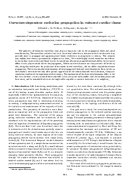
2.3. Obstacles and Drug Treatment. Obstacles in the cardiac cell sheets were made by precise cuts in the central area of cultured monolayer with 27G syringe needle controlled by micro manipulator under the phase-contrast microscope (IX-71; Olympus), just before the observation. In order to create the narrow isthmus, the cut line was briefly interrupted by elevating the needle, leaving small non cut area in the center (fig. la). For the modeling of spiral wave formation on a sharp feature, one-sided line was cut (fig. lb). In order to control the excitability and the restitution, either Lidocaine or
Electrical stimulation
(a)
(b)
Electrical stimulation
27 mm
Cutting line with syringe needle
Fig. 1. (a) - Narrow isthmus model, (b) formation model
Spiral wave
Nifekalant, or the mixture of thereof were added to the incubation solution. Lidocaine was added to the final concentrations ranging from 0.2 to 1.0 mM. Nifekalant was added in concentration range from 1.0 to 10/¿M.
2.4. Experimental Protocol. Bipolar stimulating electrode was positioned at the rim of the myocyte layers in order to minimize the damage to the studied area in the center of the layer. Trains of pulses were applied at 2.0 V and 20 ms, at least 10 s with a progressive decrease of stimulation cycle length (CL) from 1.0 s to 300 ms until the capture failure. In the case when excitation wave could not be initiated with the stimulation CL of Is, the stimulation CL increased until 2.0 s or the voltage of stimulation increased until 4.0 V.
2.5. Computational model. Wave front propagation through an isthmus was simulated using a continuous isotropic cardiac sheet model discretized on a rectangular mesh with no-flux boundary conditions [19]. The typical mesh size was 1.5 x 1.5 mm2 with dx = 5 / /111. The equation governing the transmembrane potential (Vm, mV) at each node was
dvr
dt
m = DV2Vm
-^iori Cm,
(i)
where Cm is the membrane capacitance (1.0 /¿F • cm"2), D is the diffusion coefficient (0.9cm2-s_1), I-,on is the total current flowing through the membrane (/¿A • cm"2), and V2 is the 2D-spatial Laplacian (cm"2). Equation (1) was integrated using a forward Euler method with a time step of dt = 0.04 /ts. The Laplacian was evaluated explicitly using a 9-point second difference approximation. The membrane current kinetics of I-,on are described by Hodgkin-Huxley type equations with the Drouhard-Roberge [20] formulation of the inward sodium current and the Beeler-Reuter [21] formulations of the slow inward current (Is), time-independent potassium current (//vi), and time-activated outward current (Ix 1). To approximate the action potential duration of rat neonatal myocytes the time constants of Ca2+ cur-
(a)
(b)
(c)
(d)
0 0.1 0.2 ^Йг о.з
0 1 > W 0.1 0.2
0 V/ 0.l| 0.2 к. .м 0.3
0 # 0.1 0.3
(f)
>/4
<N
(g)
(h)
E E
t (s)
Fig. 2. Propagation pattern through the narrow isthmus on the cultured cardic tissue, (f)-(h) - Time-space plot of the wave propagation beyond the isthmus. Experimental parameters are followings
Для дальнейшего прочтения статьи необходимо приобрести полный текст. Статьи высылаются в формате PDF на указанную при оплате почту. Время доставки составляет менее 10 минут. Стоимость одной статьи — 150 рублей.