m
EHOOPrÄHH^ECRAa XHMH3, 2014, moM 40, № 1, c. 20-30
DISORDERED BINDING REGIONS OF EWING's SARCOMA
FUSION PROTEINS
© 2014 t. R. Todorova#
Institute of Biophysics and Biomedical Engineering, Bulgarian Academy of Sciences, Sofia, 1113 Bulgaria
Received April 8, 2013; in final form, July 21, 2013
A relationship was found between the Amino acid (AA) composition, Intrinsic Protein Disorder (IPD) and Protein Binding Regions (PBRs) of the functional regions of Ewing's sarcoma protein (EWS) and oncogenic EWS fusion proteins (EFPs). EWS has high IPD and 64% predicted Disordered Binding Regions (DBRs) by ANCHOR. The native Transcription Factors, fused to EWS Activation Domain (EAD) in EFPs, show high DBRs in N-terminal domain and relatively low in C-terminal domain. EFPs oncogenic function is related to IPD and PBRs probabilities, high around breakpoint and decreased in the fused Transcription Factor. The increased IPD in EAD around (AA 82), and the small RBRs around (AAs (50—60) and 100) are consistent with the reported physical interactions with RNA Polymerase II subunits. The AAs (228—264) of EWS, interacting with ZFM1 (SF1), correspond to two peaks of DBRs by Anchor and high IPD by IUPred. The IQ domain of EAD (AAs 258-280) that is phosphorylated by PKC and interacts with calmodulin, has high IPD and DBRs probability. The Ser266, conserved site of PKC phosphorylation, is situated in DBR and IPD region with about 100% probability. The small PBRs found in the EAD correspond to important physical protein-protein interactions, confirmed by experimental data. Thus regions of EWS and EFPs, included in functional interactions with other partners, are enriched of Protein Binding Regions by ANCHOR. The development of IPD- and PBRs-related, EWS-FLIl-directed specific therapies will help the design of antitumor agents against ESFT because of high patient mortality in cases of meta-static disease.
Keywords: Ewing's sarcoma protein, EWS oncogenic fusions, function-structure-disorder, Predictors, Targeted inhibition, Intrinsically disordered proteins, Disordered Binding Regions
DOI: 10.7868/S0132342314010114
1. INTRODUCTION 1.1. Intrinsic Disorder
Proteins conditionally can be separated in two big groups-the group of ordered proteins and the group of Intrinsically Disordered Proteins (IDPs). IDPs are characterized by their properties (conformation ensemble, aggregation, protein translational modifications, binding diversity, coupled folding-folding) and application (protein design, new target for drug design, structure determination) [1]. Proteins do not have necessarily to be tightly folded into specific 3D
Abbreviations: EWS — Ewing's Sarcoma Protein; CTD, NTD — C- and N-Terminal Domain; ID — Intrinsic Disorder; ISD — Intrinsic Structural Disorder; IPD — Intrinsic Protein Disorder; IDPs — Intrinsically Disordered Proteins; DBRs — Disordered Binding Regions; EAD — EWS Activation Domain; AA — Amino Acid; TAD — Transcription Activation Domain; PBR — Protein Binding Regions; Pol II — RNA Polymerase II; TF — Transcription Factor; ESFT — Ewing's Sarcoma Family of Tumours.
# Corresponding author (phone: +359-2-979-2103; e-mail: todorova@bio21.bas.bg).
structures to be functional. Disordered functional proteins can be described by various techniques including X-ray crystallography, NMR, circular dichroism, hydrodynamic methods, proteolytic sensitivity, etc. "Intrinsically disordered proteins" or "native disordered" proteins have important biological functions. Intrinsically disordered proteins (and regions) lack stable structure and are linked to the function in signaling, regulation and control. Proteins, associated with human diseases, such as cancer, are enriched in intrinsic disorder: they enter in high-specificity-low-affinity interactions by one-to-many binding mode. Thus a single IDP/IDR binds to multiple structurally diverse partners, accomplished by their plasticity [2]. Disordered protein function relies upon intrinsic movement to participate in multiprotein complexes like transcription. The characterization and role of disordered proteins in human diseases represents a novel intersection of biochemistry and pathology.
EWS
EWS-FLI EWS-ATF1
SYGQ
SYGQ
SYGQ
RGG RRM RGG Zn RGG
DNA-BD
Pro
ß Q2
bZIP
EWS-ZSG
SYGQ
A/T
Zn Finger
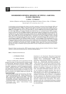
Fig. 1. Functional regions of proteins EWS, EWS-FLI, EWS-ATF1 and EWS-ZSG. Domain structure of wild-type EWS: SYGQ is serine-tyrosine-glycine-glutamine rich transactivation region; RGG is arginine-glycine-glycine rich regions; RRM is RNA-recognition motif; Zn is putative zinc finger. Domain structure of EWS-FLI: DNA-BD is DNA binding domain; Pro is proline-rich activation domain. Domain structure of EWS-ATF1: P domain contains a critical motif DLSSD; Q2 is a glutamine-rich constitutive activation domain; bZIP is a dimerization domain. Domain structure of EWS-ZSG: A/T is A-T hook DNA binding motif; Zn finger is Cys2-His2 zinc finger.
1.2. EWS-Associated Chromosome Translocations
The genetic aberrations leading to cancer are related to a high intrinsic structural disorder (ISD), enabling fusion proteins to evade cellular surveillance mechanisms that eliminate misfolded proteins. The fusion of a DNA-binding element to a transactivator domain results in an aberrant transcription factor that causes severe misregulation of transcription [3].
Ewing's Sarcoma Oncogene (ews) on chromosome 22q12 is encoding a RNA-binding protein that is a target of tumor-specific chromosomal translocations in different sarcomas in childhood with unclear origin and poor prognosis, including Ewing sarcoma tumor, Myxoid liposarcoma, Malignant melanoma of soft parts, Desmoplastic small round cell tumor and others.
1.3. Purpose of the Study
There are some experimental findings about the functional regions of EWS protein and its oncogenic fusions (Fig. 1). Predictors of Intrinsic Protein Disorder (IPD) and Protein Binding Regions (PBRs) are also available. This study attempts to unite experimental data with protein disorder predictions of proteins with known primary structure, and to check for a relationship between their amino acid (AA) sequence, IPD, PBRs and function. The results could help in the design of antitumor agents against EWS related childhood sarcomas.
2. EXPERIMENTAL SECTION
The Predictors IUPred [4], GlobPlot2 [5], DisEMBL [6], FoldIndex [7], RONN [8], PONDR [9] were used to estimate the protein disorder of native EWS and EWS oncogenic fusion proteins (EFPs) EWS-FLI1, EWS-ATF1, EWS-ZSG. Different isoforms of EWS oncogenic fusion proteins were used in IPD predictions. The Protein Binding Regions (PBRs) for the na-
tive EWS and its fusion proteins were predicted by ANCHOR [10] and compared to IPD by IUPred [4]. The values of IPD above 0.5 are considered disordered
[3]. The analysis of functional regions included reported experimental data, protein databases, and amino acid sequences for protein isoforms from NCBI (www.ncbi.nlm.nih.gov). Results of IPD and PBRs isoform predictions by IUPred and ANCHOR that are representative for all other protein isoforms, are reported in this paper.
3. RESULTS AND DISCUSSION
The fusion of EWS protooncogene with transcription factors Flil, ATF1 and EWS-ZSG creates oncogenic EWS fusion proteins (EFPs), which are potent transcriptional activators that combine the highly repetitive, disordered EWS activation domain (EAD) and the DNA-binding region of the fusion partner. Their trans-activation function is located within the N-terminal 86 amino acids of EWS [11]. Thus, EFPs promote abnormal cellular growth due to transcription deregulation of target genes [3].
The predictions of IPD for native EWS and its on-cogenic fusion proteins (EFPs) with known sequence are obtained by different Predictors [4—9] and show similar results (Table 1). Comparing, EFPs show similar IPD in the N-terminal domain (NTD) (AA 1-264, EAD) and different IPD in the C-termi-nal domain (CTD) from EWS, and between the different oncogenic fusions (Figs. 2a-2d). The longest region of EAD, free of Y motifs (AA 132-156), and the IQ domain (a Y-free region flanked by two Y-boxes (AA 258-280)) are disordered. EWS functional regions RGG1, RGG2 and RGG3 are predominantly disordered. Here are shown the IUPred
[4] predictions of intrinsic disorder (Long disorder) for EWS protein and the fusion proteins EWS-FLI1, EWS-ATF1 and EWS-ZSG, in relation to their Pro-
1.2
1.0
0.8
£ 0.6 HH
0.4 0.2
0
EWS isoform 2
/
(a)
J.
■I—I.
1 42 83 124 165 206 247 288 329 370 411 452 493 534 575 616
Residue
-1 IUPred ——— 2 RONN-3 (grey) DisEMBL-coils
1.2
1.0
0.8
g 0.6 HH
0.4 0.2
0
EWS Flil type 1
>
^/V^H
(b)
.........
1 30 59 88 117 146 175 204 233 262 291 320 349 378 407 436 465
Residue
-1 IUPred ------2 RONN-3 (grey) DisEMBL-coils
EWS-ATF1 type 2
0
1 27 53 79 105 131 157 183 209 235 261 287 313 339 365 391 417
Residue
-1 IUPred - 2 RONN -3 (grey) DisEMBL-coils
1.2
1.0
0.8
g 0.6 HH
0.4 0.2
0
H» A
EWS-ZSG long B isoform
(d)
y—I—I—I—I—I—I—I—I—I—I—I.
1 39 77 115 153 191 229 267 305 343 381 419 457 495 533 571 609
Residue
-1 IUPred ------2 RONN-3 (grey) DisEMBL-coils
Fig. 2. Comparison of IPD probability for native EWS native and its oncogenic fusion proteins by different Predictors. (a) IPD probability for EWS isoform 2 (656 AA). (b) IPD probability for EWS-Fli1 type 1 (476 AA). (c) IPD probability for EWS-ATF1 type 2 (432 AA). (d) IPD probability for EWS-ZSG long B isoform (609 AA).
1 43 85 127 169 211 253 295 337 379 421 463 505 547 589 631 673
Amino acid residue
-EWS isoform 2 -----EWS-Fii1 type 1 -----EWS-Flil type 2
EWSR1/FLI1 type 1--EWS-ATF1 type 2 — EWS-ATF1 type 1
----EWS-ZSG long B isoform ......EWS-ZSG long Aisoform ------ EWS-ZSG Short isoform
1 34 67 100 133 166 199 232 265 298 331 364 397 430 463 496 529 562 595 628
Residue
■ EWS-Fli1 type 1-
EWS-ATF1 type 2
(grey) EWS-ZSG long B
EWS isoform 2
Fig. 3. Disorder prediction score by IUPred and P
Для дальнейшего прочтения статьи необходимо приобрести полный текст. Статьи высылаются в формате PDF на указанную при оплате почту. Время доставки составляет менее 10 минут. Стоимость одной статьи — 150 рублей.
