БИОЛОГИЧЕСКИЕ МЕМБРАНЫ, 2012, том 29, № 1-2, с. 6-12
= ОБЗОРЫ
УДК 577.25
DIVERSITY OF PHOSPHOINOSITIDE SIGNALING
© 2012 B. Hille
Department of Physiology and Biophysics, University of Washington School of Medicine, Campus Box 357290, Seattle, WA 98195-7290, USA; Tel: 206-543-8639; Fax: 206-685-0619;
e-mail: hille@u.washington.edu Received 02.08.2011
Phosphoinositide phospholipids are key membrane signals that activate intrinsic membrane proteins and are loose anchor points for peripheral membrane proteins. They are also precursors for numerous second messengers. This essay reviews historical development, current opinions, and future questions about phospho-inositide signaling.
Keywords: phosphoinositide phospholipids, second messengers, membrane, signaling.
I dedicate this essay to Professor Boris Izraelevitch Khodorov with affection on the occasion of his 90th birthday, celebrating more than 70 years of extraordinary scientific productivity. Professor Khodorov has been a dear friend and a close scientific competitor since I was a Ph.D. student in the early 1960s. Some overlapping topics our laboratories have pursued in parallel include: (i) voltage clamp of nodes of Ranvier of myelinated nerve fibers; (ii) the interrelation ofvolt-age-gated Na+ channel inactivation with the actions of local anesthetics and antiarrhythmics; (iii) use-dependent (cumulative) open-channel block by these molecules; (iv) modification of many channel properties by lipid-soluble toxins including batrachotoxin and ver-atridine; (v) modification of Na+ channel gating by scorpion toxins; and (vi) regulation of cytoplasmic calcium by uptake into mitochondria. Accompanying the great similarity of our intellectual interests, Khodorov and I have constantly exchanged letters, reprints, ideas, and scientific challenges. This valuable exchange continues even now. I admire and salute his unabated intellectual energy, scientific insights, and enthusiasm.
This tutorial review essay focuses on signaling by the phosphoinositide phospholipids of cell membranes. These lipids and their metabolic products serve both as localization signals and cofactors for proteins and as second messengers for receptor signaling. Like other second messengers, the phosphoinositides and their downstream products are minority molecules with short metabolic half lives. In the cell, they are important more for their information content than for their contributions to the architecture of lipid bilayers. They participate in many physiological responses to hormones, neurotransmitters, and growth factors. Our understanding of phosphoinositide signaling has moved forward in big steps in each of the last six decades yet is still likely to be incomplete today. New ideas continue to emerge. Here, brief descriptions of
current concepts are combined with a review of some classical historical steps.
THE PHOSPHOINOSITIDES, RARE ACIDIC MEMBRANE LIPIDS
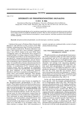
Phosphoinositides are phospholipids found primarily in the cytoplasmic leaflet (i) of the eukaryotic plasma membrane and (ii) of all eukaryotic organellar membranes. Most enzymes of phosphoinositide metabolism are cytoplasmic enzymes. The simplest, most abundant, but perhaps least interesting phosphoi-nositide is phosphatidylinositol (PI) itself (Fig. 1A). Like most other phospholipids, PI combines membrane-bound ,s«-1,2-diacylglycerol linked by a phos-phodiester phosphate to a hydrophilic head group, in this case the cyclic sugar-like myo-inositol. Seven, more interesting and much less abundant [1, 2] phos-phoinositides are formed from PI by cytoplasmic lipid kinases that phosphorylate the inositol 3, 4, or 5 positions in every combination (Fig. 1B) [3, 4].
In the cell, these phosphorylated lipids are membrane-specific, with, for example, phosphatidylinositol 4,5-bisphosphate (PI(4,5)P2) (Fig. 1B) being the signature phosphoinositide of the plasma membrane, phosphatidylinositol 4-phosphate (PI(4)P) being the signature phosphoinositide of Golgi, transport vesicles, and secretory vesicles, and PI(3,4)P2, PI(3)P, and PI(3,5)P2 being the signature phosphoinositides of various endosomal compartments . During membrane trafficking, as one compartment delivers membrane to another, the phosphorylation of the phosphoinositides is reworked to match the destination compartment within seconds by recruited specific lipid kinases or phosphatases. PI(4,5)P2 is highly acidic, carrying about 4 negative charges at physiological pH. These phosphorylated phosphoinositides account for only a few percent of all the acidic phospholipid of eukaryot-
Phosphoinositide backbone
glycerol phosphate o. inositol H°
OH
O D5
B
All phosphoinositides
Rapidly inter-converted membrane ZIP-code PI lipids
Lipid Name PI
PI(3)P
PI(4)P
PI(5)P
PI(3,4)P2
PI(3,5)P2
PI(4,5)P2
PI(3,4,5)P3
often called
Inositol phosphorylation
D3 D4 D5
H H H
P H H
H P H
H H P
P P H
P H P
H P P
P P P
'PIP,
O 5
D4
Fig. 1. The structure of phosphoinositides. A, A generic phosphoinositide showing the D3, D4, and D5 positions on the inositol head group that can be phosphorylated. When none is phosphorylated, this is the root compound, PI. B, Nomenclature for the seven phosphorylated forms.
ic cells [1]. They are low abundance lipids but with great significance.
CANONICAL RECEPTOR SIGNALING THROUGH PHOSPHOLIPASE C
In mammals, about 50 different G-protein coupled receptors (GPCRs) couple via the G proteins Gq and G11 to phospholipase Cp (PLCp) (Fig. 2A). Typical physiological examples of this "hormone-sensitive pathway" are activation of salivary secretion or of pancreatic exocrine secretion by acetylcholine acting on M1 muscarinic receptors (M1R). Growth factors that act through tyrosine-kinase receptors engage several diverging signaling pathways, one of which involves another phospholipase, PLCy. The canonical downstream result is the same for PLCp and PLCy, namely the Ca2+-dependent PLC cleaves its substrate PI(4,5)P2 into the hydrophobic, membrane-associated diacylglycerol (DAG) moiety and soluble cytoplasmic inositol 1,4,5-trisphosphate (IP3) (Figs. 2A and 3). Both molecules are potent second messengers that make more signals. IP3 opens IP3 receptor channels on the endoplasmic reticulum to release Ca2+ from intracellular stores. The Ca2+ rise feeds back positively on PLC activity and also stimulates many other Ca2+-de-pendent processes including exocytosis. DAG activates protein kinase C (PKC) and other proteins that have a C1 domain that recognizes DAG. Still only incompletely known is the fate of the IP3. Its cytoplasmic lifetime is perhaps ~15 s, but what happens to it then? Classically, phosphoinositol phosphatases were shown to make inositolbisphosphate, inositolmono-
phosphate, and finally free inositol for reuse. However, there is increasing discussion of polyphosphoinositol kinases that make highly phosphorylated forms up to IP8 (with several positions pyro--phosphorylated). When does IP3 become dephosphorylated and when does it become polyphosphorylated? Some of the higher phosphoinositols are abundant (hundreds of micromolar) and have actions on or are even necessary for several cellular processes [5, 6]. Perhaps in ten years we will have better perspective on their significance. Other consequences of Gq activation include stimulation of MAP kinases and formation of many other lipid messengers like arachidonic acid, phospha-tidic acid, prostaglandins, prostacyclin, thrombox-anes, and leukotrienes. Thus, an amazingly rich diversity of cytoplasmic and membrane messages is derived from PI(4,5)P2 upon stimulation of PLCp or PLCy by hormones, neurotransmitters, and growth factors.
For the Khodorov laboratory and ours, the release of calcium by IP3 and the activation of PKC have been especially interesting consequences of this signaling from phosphoinositide cleavage.
SELECTED KEY HISTORICAL DISCOVERIES IN RECEPTOR-MEDIATED PLC ACTIVATION
A lipid connection in hormone action was prominently revealed by Mabel and Lowell Hokin [7, 8]. In a long series of groundbreaking papers they showed clearly that muscarinic stimulation of several secretory tissues greatly speeded the rate of incorporation of 32P phosphate into phosphoinositide lipid while the tissue was secreting its peptide product. They hypothesized
receptor + agonist
A
B
Inhibition
Recovery
PI 4-kinase
PI(4)P
А
PI(4)P 5-kinase
Í:''""^TPI(4,5)P
P P
PI(4)P 4-phosphatase
PI(4,5)P2 5-phosphatase
Ion channel
Fig. 2. Chemical reaction scheme for the steps of PI(4,5)P2 metabolism. A, Receptor signaling to PLCp and cleavage of PI(4,5)P2. B, Resynthesis of PI(4,5)P2 from PI. The lipid kinase reactions are dynamically opposed by lipid phosphatases. PI(4,5)P2 supports the activity of many ion channels.
hydrophobic
membrane ) ^
arachidonic stearic ) \ fatty acid chains in
acid 1 , J < j < acid ) \ innerlipidleafletof
j S plasmamembrane
inositol
O
oP-o-
O-
Phosphatidylinositol 4,5-bisphosphate (PIP2)
cytoplasm
Inositol l,4,5-trisphosphate (IP3)
Fig. 3. Calcium-requiring phosphoinositol-specific PLC cleaves PI(4,5)P2 to yield two versatile second messengers DAG and IP3.
that muscarinic activation directly stimulates synthesis of phosphoinositide (not correct) and that phosphoinositide is a mediator of "active transport." At that time, the concept of exocytosis was not current, and "active transport" might refer equally to secretion of proteins, ions, and fluids. By the early 1970s, Robert Michell [9] developed the concept that many receptors stimulate a calcium-dependent enzyme PLC (called carefully in the British literature polyphospho-inositide phosphodiesterase) that cleaves the phos-phoinositide PI(4,5)P2 into DAG and "inositol products". The PI-specific and calcium-sensitive PLCs were first isolated and studied by the lab of Philip Ma-jerus [10], and their full diversity and regulation was
elaborated by Sue Goo Rhee (summarized by Rhee et al., [11]). The enzymolo
Для дальнейшего прочтения статьи необходимо приобрести полный текст. Статьи высылаются в формате PDF на указанную при оплате почту. Время доставки составляет менее 10 минут. Стоимость одной статьи — 150 рублей.