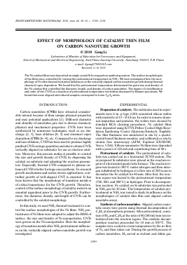
НЕОРГАНИЧЕСКИЕ МАТЕРИАЛЫ, 2010, том 46, № 10, с. 1190-1194
EFFECT OF MORPHOLOGY OF CATALYST THIN FILM ON CARBON NANOTUBE GROWTH © 2010 Gang Li
Laboratory of Ministry of Education for Conveyance and Equipment, School of Mechanical and Electrical Engineering, East China Jiaotong University, Nanchang 330013, P.R. China
e-mail: ligang0794@163.com Received 11.10.2009
The Ni catalyst films were deposited on single crystal Si by magnetron sputtering system. The surface morphologies of the films were controlled by varying the pretreatment temperature in NH3. We have investigated how the morphology of Ni after thermal treatment influences on the vertically aligned carbon nanotubes growth during thermal chemical vapor deposition. We found that the pretreatment temperature determined the grain size and density of the Ni catalyst that controlled the diameter, length, and density of carbon nanotubes. The degree of crystallization and order of the CNTs as a function of pretreatment temperature was further discussed by Raman spectrums. We found that more aligned and ordered samples correspond to lower ID/IG ratios.
INTRODUCTION
Carbon nanotubes (CNTS) have attracted considerable interest because of their unique physical properties and may potential applications [1]. Different diameter and chirality of nanotubes give rise to diverse chemical, physical and mechanical properties. CNTs have been synthesized by numerous techniques, such as arc discharge [2, 3], laser ablation [4, 5] and chemical vapor deposition (CVD) [6—8], etc. Compared to arc discharge and laser ablation, CVD has been used to produce highly purified CNTs in large quantities and also to obtain CNTs vertically aligned on substrates for use as electron emitters. Moreover, this process makes it possible to control the size and growth density of CNTs by dispersing the catalyst on substrate and adjusting the reaction parameters. Especially, thermal CVD compared to plasma enhanced CVD is better for large area synthesis. To research growth mechanism and realize device applications, controlled growth of well-aligned CNT is essential. It has been known that the morphology of transition metals is of critical importance for the CNTs growth. Therefore, control of the surface morphology of catalytic metal is an essential ingredient prior to the CNTs growth. Furthermore, the diameter, density, and length of CNTs can be controlled by the catalyst morphology.
In this study, we used NH3 thermal treatment to control the surface morphologies of the Ni films. NH3 pretreatment of Ni films were adopted to adjust the RMS of surface, the size and density of Ni nanoparticles. CNTs were grown on the Ni nanoparticles. How the morphology oftransition metals after NH3 pretreatment influences on the vertically aligned carbon nanotube growth was investigated.
EXPERIMENTAL
Preparation of catalysts. The substrates used in experiments were 6-in. p-type (100) orientated silicon wafers with resistivity of15—25 fi cm. In order to remove chemical impurities and particles, the wafers were cleaned by standard RCA cleaning procedures. Ni catalyst films were deposited using K575X Peltier Cooled High Resolution Sputtering Coater (Quorum/Emitech, English). The film thickness was monitored in situ by a quartz-crystal based thickness monitor, and calibrated ex situ by atomic fore microscopy (AFM, Explorer EX300500, Veeco, USA). Fifteen nanometer Ni films were deposited with a power of 120 mA and a sputtering time of 40 s.
Pretreatment of catalysts. The pretreatment of catalysts was carried out in a horizontal TCVD system. The as-prepared Si substrates were placed in the reaction region of a horizontal quartz tube furnace. The reaction region was heated to 580°C under nitrogen and then nitrogen substituted by hydrogen at a flow rate of 200 sccm to deoxidize the Fe catalyst for 40 min. After that, the reaction region was heated to the pretreatment temperature (700, 800, and 900°C) in hydrogen. Prior to decomposition reaction, Fe catalyst on Si substrates was pretreated in NH3 gas for 20 min. The temperature of catalyst pretreatment in NH3 was varied to study its influence on the morphologies of catalyst thin film and aligned carbon nanotube arrays.
Synthesis of carbon nanotubes. Aligned carbon nano-tube arrays were grown using thermal decomposition of C2H2 at atmospheric pressure. After pretreatment, a mixture ofH2 and C2H2 at the ratio of40/200 ml/min was introduced into the reaction region. The catalytic decomposition reaction proceeded for 10 min at 850°C. The samples were cooled to room temperature by the in-flow of N2 and then taken out. During the growth process of carbon nanotubes, H2 served as etchant and dilute gas.
(a) (b)
(d)
20
й
t/T
s
10
800 900 1000
Temperature, °C
Fig. 1. AFM images of surface morphology for Ni films after pretreatment for 15 min: (a) 800°C, (b) 900°C, (c) 1000°C, and (d) height RMS of roughness as a function of the pretreatment temperature.
The purities of the gas employed in the experiment are all higher than 99.5%.
Characterization. The surface morphologies of nickel thin films were examined by atomic force microscopy (using a non-contact mode of atomic force microscopy (AFM). The length, diameter and density distributions of CNTs were observed by field emission scanning electron microscope (FESEM, JEOL JSM-7001F, Japan). The crystallinity of CNTs was characterized by Raman spectroscope (JY, T64000, France) using 514.5 nm Ar excitation.
RESULTS AND DISCUSSION
Effects of NH3 plasma pretreatment. We observed the effect of NH3 gas on the formation of Ni nanoparticle during pretreatment. Figures 1a, 1b, and 1c are the AFM images of surface morphology of Ni catalytic thin fims with 15 nm in thickness etched at different pretreatment temperature in NH3 ambient for 10 min. The corresponding temperature is 800, 900, and 1000°C in images Fig. 1a, 1b, and 1c, respectively. We observed that the Ni thin was transformed into nanoparticles. These nanopar-ticles could act as the nucleation sites for growth of carbon nanotubes. As the pretreatment temperature increased from 700 to 900°C, an average diameter of Ni
particles increased and the density of particles deceased. These results suggesting that Ni film agglomerated to become large particles due to the increase of mobility of nanoparticles on the substrate surface at higher temperatures during pretreatment [9, 10]. It was believed that diffusion, nucleation and etching would occur during NH3 plasma pretreatment. The NH3 plasma pretreatment also provided cleaning effect which would help growth of carbon nanotubes. And both small nanoparticles and partially thin films were etched and then removed by NH3 gas. The RMS surface roughness of the Ni thin films changed from 6.14 to 22.68 nm as pretreatment temperature increased from 800 to 1000°C, as shown in Fig. 1d.
Growth and properties of carbon nanotubes. Figures 2a, 2b and 2c show the SEM images of aligned carbon nan-otubes grown on the substrate with Ni particles that were pretreated with the previously described conditions. The edges are peeled off using a razor, in order to be visualized. For the case of NH3 pretreatment with 800°C, the nanotubes are oriented perpendicular to the surface of the substrate due to high density of Ni nanopartilces as shown in Fig. 2a. When NH3 pretreatment temperature of increased up to 900°C, because of a low particle density, the nanotube density is also low and the nanotubes are not strictly oriented vertically as shown Fig. 2b. In
(a)
(b)
800 900 1000
The pretreatrent temperature, °C
Fig. 2. SEM images of CNTs grown on nanoparticles corresponding to Fig. 1, where insets are High-resolution SEM images of the CNTs, respectively.
particular, we observed that at the pretreatment temperature of1000°C, (I) the CNT lengths were relatively short compared to the CNTs grown at other conditions and (II) the CNTs were no longer aligned due to the lower Ni particle density as shown Fig. 2c. So from Fig. 2, the density of CNTs decreased from 82 to 216 ^m-2, and CNTs showed worse alignment as the pretreatment temperature increased from 900 to 1000°C. From the inset images, the ranges ofthe diameter ofCNTs increase from 50-100 to 100-250 nm with increasing the pretreatment temperature. The CNT length decrease with the RMS ofsur-face morphology, while the CNTs diameter increase with the RMS, as summarized in Fig. 2d. As we know, catalysts played a critical role in the synthesis of carbon nan-otbues. Their densities and sizes controlled the morphologies of CNT arrays. It strongly suggests that the formation mechanism of the vertically aligned CNTs is the steric hindrance between nanotubes, which is invokes by the high number density of Ni grains.
Raman spectroscopy, a powerful technique, is widely used to characterize the carbon materials. Crystalline
graphite belongs to the space group the zone-center optic phonon modes can be decomposed into the following irreducible representations
ropt ^ 2E2g + Eu + Au + 2B2g. (1)
Among which only the two E2g modes are Raman active [11], the E1u and theA2u are infrared active, and the B2g modes are optically inactive. Raman spectra of CNTs grown on grown on nanoparticles corresponding to Fig. 1 are shown in Fig. 3. The information collected from the Raman spectroscopy investigation included the Raman-peak intensities for D and G peaks, that is ID and IG peaks, the ID/IG ratios and the position for the IG and IG as shown in Table.
From Fig. 3, all the CNTs apparently consist of two peaks near 1350 (D -band) and 1580 cm-1 (G -band) similar to that of crystalline graphite. The G-band corresponds to a phonon high-frequency E2g first order mode of graphite and is related to the vibration of sp2-bonded carbon atoms in a 2-dimenstional hexagonal lattice, such as in graphite layer [12, 13]. On the other hand, the D-band is associated with vibrations of carbon atoms with dangling bonds in plane termin
Для дальнейшего прочтения статьи необходимо приобрести полный текст. Статьи высылаются в формате PDF на указанную при оплате почту. Время доставки составляет менее 10 минут. Стоимость одной статьи — 150 рублей.