КОЛЛОИДНЫЙ ЖУРНАЛ, 2010, том 72, № 5, с. 714-720
УДК 541.183
EFFECTS OF SURFACTANT/WATER RATIO AND DYE AMOUNT ON THE FLUORESCENT SILICA NANOPARTICLES © 2010 г. Yanhua Sun1, 3, Xian Wang1, Jiangjiexing Wu2, Yan Fu1, Jinli Zhang1, Hao Li1, Wei Li2
1Key Laboratory of Systems Bioengineering and 2Key Laboratory for Green Chemical Technology, Ministry of Education, School of Chemical Engineering & Technology, Tianjin University,
Tianjin 300072, P. R. China 3School of Basic Medical Science, Tianjin Medical University Tianjin 300070, P. R. China Поступила в редацию 18.08.2009 г.
Effects of surfactant/water volume ratios and dye amounts on the properties of micelles and fluorescence silica nanoparticles were studied in microemulsions containing nonionic surfactant Triton X-100, hexanol as co-surfactant, cyclohexane as organic solvent, and metal organic dye (tris(2,2'-bipyridyl)dichlororuthenium) via fluorescence probe technique, TEM, and XPS. Fluorescence probe measurements show that the micelle microenvironment becomes stable at the surfactant/water volume ratio higher than 3.5 and the incubation time longer than 10 h. The data suggest that the silica shell, which is formed at the surfactant/water ratio of 3.5, yields highly efficient protection of dye molecules against the e-beam irradiation and result in high pho-tostability of fluorescent silica. We pioneered the partial localization of dye molecules on the surface of dye-doped silica and found that an increase of dye amounts beyond a threshold in the microemulsion cannot enhance the fluorescence intensity of dye-doped nanoparticles. These results are of significant importance for optimizing the synthesis of fluorescent nanoparticles with high photostability and low cost.
INTRODUCTION
Nanoparticle synthesis in microemulsions has attracted increasing attention due to its importance for many applications including catalysts [1], medicine [2], nanofluids [3] and especially fluorescent bioassays [4—8]. Dye-doped fluorescent silica nanoparticles have been synthesized by using water-in-oil microemulsions with the dopant ofvarious dye compounds such as tris(2,2'-bi-pyridyl)dichlororuthenium(II) [9, 10], tris(2,2'-bipy-ridyl) osmium(II) bis(hexafluorophosphate) [11], fluorescein [12], and dextran-modified tetramethylrhodamine [13]. The studies attempting to improve the performance of dye-doped nanoparticles in antigen-antibody detection, bioimaging and other high-throughput bioassays have reported that microemulsion parameters including the nature of surfactants and cosurfactants, hydrolysis reagents, solvents and the water content can significantly influence the size and properties of fluorescent nanoparticles [14, 15]. For example, Zielinska et al. have reported that the chemistry ofboth the surfactant and co-surfactant affect the shape and size of micelles in micro-emulsions containing the surfactant N-alkyl-N-methyl-gluconamides and certain alcohols as cosurfactants [16]. Tan and coworkers have found that tris(2,2'-bipy-ridyl)dichlororuthenium(II)-doped silica nanoparticles synthesized at different water to surfactant molar ratios in Triton X-100/cyclohexane/hexanol system had a large size distribution from 69 nm to 178 nm [17]. Generally, the dye dopants are expensive and comprise, to a large extent, the cost of fluorescent nanoparticles. However,
no reports, which would describe how to synthesize fluorescent nanoparticles with the lowest possible dye amount, have been published yet.
The reverse micellar interfaces confine dynamic aggregates undergoing collision, coalescence and dissociation in water-in-oil microemulsions [18]. It is necessary, therefore, to study whether the dye molecules locate on the surface or inside the nanoparticles. The answer to this question is important for optimizing the synthesis of fluorescent nanoparticles with high photo-stability and low cost.
In this article, we used fluorescence probe techniques to investigate the effects ofsurfactant/water volume ratios on the micelle microenvironment for microemulsions comprising the nonionic surfactant Triton X-100, hex-anol as cosurfactant, cyclohexane as organic solvent, and a metal organic dye (tris(2,2'-bipyridyl)dichlororutheni-um). The photostability and localization of dye molecules in the dye-doped silica nanoparticles synthesized at certain surfactant/water ratios were characterized by TEM, fluorescence spectroscopy and XPS. It was shown that the silica shell formed at the surfactant/water ratio of 3.5 provide highly-efficient protection of dye molecules against the e-beam irradiation and results in high photostability of fluorescence dye-doped nanoparticles. The results suggest that an increase of dye amount beyond a threshold in the microemulsion has a negative effect on the fluorescence intensity of dye-doped nanoparticles.
MATERIALS AND METHODS 2.1. Materials
The tris(2,2'-bipyridyl)dichlororuthenium (Ru(bpy)3Cl2 ■ 6H2O, abbreviated as Ru-dye), Triton X-100 (the average molecular weight of 647), silica precursor tetraethylorthosilicate (TEOS) and the pyrene probe were purchased from Sigma-Aldrich. Cyclohex-ane, hexanol, aqueous ammonia (NH3 ■ H2O) solution (25 wt %), acetone and anhydrous ethanol were purchased from Tianjin Chemical Reagent Co., which were ofA.R grade and used without further purification.
2.2. Fluorescence probe measurements
Microemulsions were prepared at 25°C by mixing different volumes of Triton X-100 (0.70-1.97 mL), 1.8 mL hexanol, 500 ^L water, and 7.5 mL cyclohexane containing 10-5 M pyrene probe, and were incubated for different periods of time (5 min—24 h) before fluorescence measurements. Fluorescence spectra were recorded in the 350-600 nm range at 25°C, with an excitation band of pyrene at 334 nm. The fluorescence peaks at 373 nm (/1) and 384 nm (I3) were used to calculate the I1/I3 ratio that indicates the polarity of local environment.
2.3. Preparation of fluorescent dye-doped silica nanoparticles
The microemulsion was stirred at room temperature and 120 rpm for 24 h (if it is not mentioned otherwise) after mixing 1.77 mL of Triton X-100, 1.8 mL of hexanol, 7.5 mL of cyclohexane and 500 ^L H2O containing appropriate amount of Ru-dye. 100 ^L silica precursor TEOS was introduced into the above microemulsion under stirring, this step was followed by adding 60 ^L aqueous ammonia to initiate the hydrolysis of TEOS. After 24 h, the solution was demulsified by adding 10 mL acetone, and then the dye-doped silica fluorescent nanopar-ticles were separated from the demulsified solution through centrifugation (10000 rpm, 5 min) and washed by ethanol.
2.4. Characterization
The morphology of microemulsions and the synthesized dye-doped silica nanoparticles were measured using a field emission gun transmission electron microscope (TEM, Tecnai G2 F-20, Philips) at the accelerating voltage of 200 kV Fluorescence spectra were performed with Cary Eclipse fluorescence spectropho-tometer (Vrian, USA) at the excitation wavelength of 450 nm for Ru-dye. The elemental composition of the nanoparticle surfaces was determined using X-ray photo-electron spectroscopy (XPS, PHI 1600 ESCA System, Perkin-Elmer). XPS measurements were operated at a power level of300 W with a Mg Ka X-ray source. Electron binding energies were calibrated against the alkyl C 1 s emission peak at 284.6 eV.
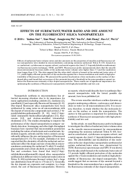
Intensity, a. u. (a)
10 8 6 4 2 0
350 400 450
500 550 600 Wavelength (nm)
10
15
20 25 Time (h)
Fig. 1. (a) Fluorescence spectra of the probe in cyclohexane; (b) plot of fluorescence intensity ratio of the first and the third vibronic peaks, I1/I3, versus the surfactant/water volume ratio in microemulsion; (c) plot of I1/I3 versus the incubation time of microemulsion.
RESULTS AND DISCUSSION
3.1. Effects of surfactant/water ratio on the micelle microenvironment
The fluorescence probe technique was utilized to study the polarity variation in the local environment during the formation of reverse micelle, using pyrene as the probe. Figure 1a shows a typical fluorescence spectrum of hydrophobic pyrene monomer in cyclohexane solvent and clearly displays the vibration structure including five distinct peaks. The intensity ratio of the first and the third
0
2
Fig. 2. TEM images of dye-doped silica nanoparticles prepared with the surfactant/water ratio of (a) 3.0 and (b) 3.5, respectively; (c) plots of relative fluorescence intensity of dye-doped silica with the surfactant/water ratio of (1) 3.0 and (2) 3.5 versus the measuring time.
vibration bands (/1//3) was proved to indicate the polarity of the probe environment [19, 20]. In order to detect the effect of surfactant/water ratio on the micelle microenvironment, fluorescence measurements were performed for microemulsions with different surfactant/water ratios incubated at 25°C for 24 h. According to the obtained fluorescence spectra, I1/I3 ratios were calculated (Fig. 1b). It is indicated that the I1/I3 value increases from 0.71 to 0.81 as the surfactant/water volume ratio increases from 1.4 to 4.0, with a transition point at the surfactant/water ratio of 3.5. When the surfactant/water ratio is larger than 3.5 the I1/I3 value changes negligibly upon further increase ofthe surfactant/water ratio. Taking into account the I1/I3 value (0.59) in the pure cyclohexane, it is reasonable to suggest that the polarity of the media surrounding the pyrene probe in the microemulsion is higher than that in cyclo-hexane, i.e., pyrene molecules are preferably located in a close proximity to the water—oil interface rather than in the bulk of organic solvent.
We further studied the dynamic variation of I1/I3 value at the surfactant/water ratio of 3.5. Figure 1c shows the plot of I1/I3 versus the incubation time of the microemulsion. It is indicated that the I1/I3 ratio depends on the microemulsion incubation time, especially during the first 10 h, when about 7.2% variation of the I1/I3 value is achieved. It is known that microemulsions are thermod
Для дальнейшего прочтения статьи необходимо приобрести полный текст. Статьи высылаются в формате PDF на указанную при оплате почту. Время доставки составляет менее 10 минут. Стоимость одной статьи — 150 рублей.