ОПТИКА И СПЕКТРОСКОПИЯ, 2012, том 112, № 5, с. 723-728
СТЕКТРОСКОПИЯ АТОМОВ И МОЛЕКУЛ
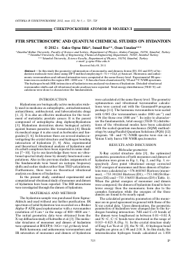
FTIR SPECTROSCOPIC AND QUANTUM CHEMICAL STUDIES ON HYDANTOIN
© 2012 n Gulce Ogruc Ildiz*, Ismail Boz**, Ozan Unsalan***
*Istanbul Kultur University, Faculty of Science and Letters, Department of Physics, Atakoy Campus, 34156, Istanbul, Turkey **Istanbul University, Faculty of Engineering, Chemical Engineering Department, 34320, Istanbul Turkey ***Istanbul University, Science Faculty, Department of Physics, 34134, Istanbul, Turkey
e-mail: g.ogruc@iku.edu.tr Received July 08, 2011
Abstract—In this study the geometry optimization of monomelic and dimeric forms (Dl, D2 and D3) of hy-dantoin molecule were done using DFT method employing 6—31++G(d, p) basis set. Harmonic and anhar-monic wavenumbers and infrared intensities were computed at the same theory level. Experimental IR spectrum was recorded in the region 400—4000 cm-1. It has also been charaterized by 1H and C NMR spectrum. The hydrogen bond (HB) interaction of hydantoin was analyzed via dimers of hydantoin. Detailed vibrational wavenumber shifts and all vibrational mode analyses were reported. Total energy distributions (TED %) calculations were done to characterize the fundamentals.
INTRODUCTION
Hydantoins are biologically active molecules widely used in medicine as antiepileptic, antischistosomal, antiarythmic, antibacterial, and tuberculostatic drugs [1, 2]. It is also an effective medication for the treatment of metastatic prostate cancer. It is the parent compound of antiepileptic drug diphenylhydantoin [3]. Hydantoin derivatives shows biological activity against human parasites like trematodeos [4]. Beside its medical usage it is also used as herbicides and fungicides [5, 6]. In literature there are several studies investigating the crystal structure and hydrogen bonding interaction of hydantoin [3, 4]. Also, experimental and theoretical vibrational analysis of hydantoin and its cobalt complexes have been reported in some studies [7-10]. Up to our knowledge there were no vibra-tional spectral study done by density functional computations. Also in the previous studies assignments of the fundamentals were based on isotopic frequency shifts and earlier studies rather than TED calculations. Furthermore, there were no theoretical vibrational analysis on dimers of hydantion.
In the present study, combined experimental and computational vibrational study of monomer and dimers of hydantoin have been reported. The HB interactions were investigated through the dimers of hydantoin.
MATERIALS AND METHOD
The hydantoin sample was purchased from Sigma-Aldrich and used without any further purification. IR spectrum of solid hydantoin was recorded on a Bruker Alpha-P ATR spectrophotometer with a wavenumber resolution of 4 cm-1 in the range of 400-4000 cm-1. The initial geometric data were obtained from the X-ray diffraction study ofSchwalbe et al. [3]. The molecular structures of monomer and dimers of hydantoin were optimized at B3LYP/6-31G++(d, p) theory level.
Both harmonic and anharmonic wavenumbers and IR intensities of monomer and dimers of hydantoin
were calculated at the same theory level. The geometry optimization and vibrational wavenumber calculations were carried out with the Gaussian09 program package [11]. The harmonic wavenumbers were scaled with 0.985 (for wavenumbers under 1800 cm-1) and 0.96 (for those over 1800 cm-1. In order to characterize the fundamentals, total energy (TED-%) distributions of the vibrational modes have been calculated with the scaled quantum mechanics (SQM) methodology by using Parallel Quantum Solutions (PQS) [12] program. 1H- and 13C-NMR spectra were run on a Varian Unity Inova 500 NMR spectrometer.
RESULTS AND DISCUSSION
Molecular geometries
X-Ray crystal structure data [3], the optimized geometric parameters of both monomer and dimers of hydantoin were given in Fig. 1, Fig. 2, and Fig. 3, respectively. Zero point vibrational energy corrected SCF energies of monomer and three dimers of hydantoin were calculated as -376.660365 Hartrees (monomer), -753.341265 Hartrees (D1), -753.340256 Hartrees (D2) and -753.336855 Hartrees (D3) (Table. 1). When the global energies of monomer and dimers were compared, the dimers of hydantoin found to have lower energy then the monomeric form due to the complex formation while the energies of the three dimers are found to be nearly equal.
The calculated geometric parameters of the monomer are in good agreement in general with those of the X-ray crystal data. Upon dimerization, the geometric parameters of hydantion were changed due to the hydrogen bonding interaction. NH and C=O bonds of the dimers were lengthened in between 0.01-0.02 Á and N-C, C-C bonds were shortened in the range of 0.013-0.025 Á (Fig. 3). In the crystal structure study of Fang-Lei Yu et al. [3], the N-H...O hydrogen bond lengths are given as 1.98 and 2.01 Á. In this study, the intermolecular hydrogen bonds calculated as 1.858
Fig. 1. X-Ray crystal structure of Hydantoin [3].
08
N1 1.478 H10
1.008 128.57
Fig. 2. Calculated bond lengths (A) and angles (degrees) of monomelic form of hydantoin.
and 1.858 A, 1.842 and 1.864 A, and 1.891 and 1.891 A for D1, D2 and D3, respectively. These results are in agreement with the crystal structure study.
The 1H-NMR spectrum of hydantoin in D2O shows two signals at 4.03 (t, J = 11.2; 2.4 Hz) and 4.65 ppm (t, J = 2.4, 11.7 Hz) attributed to CH2-NH
Table 1. SCF energies of monomer and three dimers of hydantoin
(Hartrees)1 (Hartrees)2 kcal/mol
Monomer -376.7405174 -376.660365 -236355.51
Dimer l -753.5042756 -753.341265 -472723.90
Dimer 2 -753.5028496 -753.340256 -472723.27
Dimer 3 -753.4991167 -753.336855 -472721.14
1 Calculated at DFT/6-31G++(d, p) theory level.
2 Zero point vibrational energy corrected.
and CH2-C=O protons, respectively. The 13C-NMR spectrum of the free hydantoin in D2O solution showed three signals at 177.0, 160.47 and 48.14 ppm which were assigned to C-2, C-5 and C-3, respectively [13].
Vibrational assignments
Experimental and theoretical (DFT-B3LYP/6-31G+ +(d, p)) vibrational wavenumbers of Hydantoin were given in Table. 2. Hydantoin consists of 11 atoms, which has 27 normal modes. These normal modes of the title molecule have been assigned according to the detailed motion of the individual atoms. All normal modes assigned to one of 21 types of motion (C—N, C=O, C-H and N-H stretchings; HCH, CCN, CCH, NC=O, CC=O, NCN, CNC, CNH, and NCH bendings and HNC=O, OCCH, HCNH, NCNH, CNC=O, NCCN, CNCC, and NCNC torsions) predicted by a calculational analysis.
The results obtained from the calculations show that, while the anharmonic corrections of wavenum-bers are closer to the experimental ones rather than the scaled wavenumbers, the scaled harmonic wavenum-bers of dimer forms gave the best fit to the experimental ones. The vibrational wavenumbers of both the monomeric and dimeric forms of hydantoin obtained from the DFT calculations are almost the same except the wavenumbers associated with intermolecular hydrogen bonds. To avoid complexity of huge data, calculated results for vibrational wavenumbers for monomer (as first value stands for harmonic (h) and second for anharmonic (ah)) will be given in parenthesis immediately after experimental values for the rest of the text.
v1 (NH) stretching mode for monomer was observed at 3257 cm-1 (moderate intensity) (3537-3581 cm-1). Upon dimerization for D1, H7 and O8 atoms involved in hydrogen bonding (HB) cause a downward shift in wavenumber for v1 to 3243 and 3120 cm-1, respectively. For D2, v1 shifted downward to 3255 cm-1 due to HB interaction of O8 atom. Besides, because H7 atom in D2 and O8 atom in D3 are not involved in HB wave-numbers were not shifted. v4 CH2 symmetric stretching mode was observed at 2944 cm-1 (2932-2935 cm-1) in FTIR spectrum. Because CH2 group does not contribute to HB, wavenumbers were not shifted and calculated as 2935, 2936, 2935, 2928 cm-1 for D1, D2 and D3, respectively. v5 and v6 C=O stretching modes were observed with medium IR intensity at 1774 cm-1 (1830-1819 cm-1) and strong intensity at 1696 cm-1 (1795-1786 cm-1) respectively. Wavenumbers of v5 for D1 and D2 and v6 of D3 were red shifted. v7 HCH bending mode was not observed in IR spectrum but just calculated at 1479 cm-1 -(h)-1449 cm-1 and found to be nearly same for all of the dimers because related atoms are not involved in dimerization. v8, v9 and v10 bending modes were observed with medium intensities
H10
1.096
1.096
C2 11453 1.026 JH7
1.533
1.213
09-21 C4
112.44 105.08 1.354
1.383 112.80
C3
124.59 (Nl^fW W31
;.72
? 128.7
......
O8 18
(a)
127.13
1.213
1.533
C18
H22 ''
096 H21
019
1.213
O9
H11
109.80
127.62 c4 125.80
1.096
1.537
C2
H10 1.452
1.382
107.07
(b)
1.842 O17 124.49
^fc*.......
1.230
1.031
C14
1.406
1.382
1.396
1.355
1.365 C3
112.47 105.11
C15
\
1.213 ----
1.533
126.66 1.452 C18 H21 112.90 1.096
1.096
1.097
H21
113.42 C16*^ 1 530 1.226
H17
1.008
1.214 12
C13 1.416
126.09
123.29 1.027
1.891
(c)
1.891
/
1.214
128.28
1.374
O9
127.58 106.64
127.58 c4 106.64 1.226 1.530
1.008
112.95 N1 H7
125.56
101.90 1.451
C2 113.42
1.097
H11
Fig. 3. Calculated bond lengths (A) and angles (degrees) of dimers (D) of hydantoin; (a) D1, (b) D2 and (c) D3, respectively.
Table 2. Experimental and theoretical (DFT-B3LYP/6-31G++(rf, p)) vibrational wavenumbers (cm *) of hydantoin
Modes IRexp Theoretical calculatios TED (Total Energy Distributions) (%)a
D1 D2 D3 Monomer
* VD * vd * vd * VM ** v *
v1> vnh 3257 3242 3537 3538 3537 3581 vnh(100)
3204 3255
v2> vnh 3130 3509 3509 3248 3509 3495 vnh(100)
3158 3213
v3, VCH2asym. 2973 2976 2968 2971 2932 VCH2(asym) (100)
2973
v4, VCH2sym. 2944 2936 2936 2928 2932 2935 VCH2(sym) (100)
2935
v5' vco 1774 1781 1769 1827 1830 11819 vco(81)
1749 1756 1822
v6> vco 1696 1819
Для дальнейшего прочтения статьи необходимо приобрести полный текст. Статьи высылаются в формате PDF на указанную при оплате почту. Время доставки составляет менее 10 минут. Стоимость одной статьи — 150 рублей.