ИЗВЕСТИЯ РАИ. СЕРИЯ БИОЛОГИЧЕСКАЯ, 2007, № 4, с. 406-412
= РЕГЕНЕРАЦИЯ :
УДК 57.089
IMMUNOSTIMULATION EXACERBATES THE BIOLOGICAL EFFECTS
OF CHEMICAL CARCINOGENS © 2007 V. Skourou*, Th. Keramitsoglou*, D. Koussoulakou, V. Mitashov**, S. Koussoulakos*
* University of Athens, Faculty of Biology, Department of Cell Biology and Biophysics, Athens, Greece **Kol'tsov Institute of Developmental Biology, Russian Academy of Science, 119991 Russia, Moscow, ul. Vavilova, 26
E-mail: skoussou@biol.uoa.gr Received 29.12.2006 r.
The reciprocal relation between, the high regenerative ability of various animal species and the low incidence of haphazard or experimentally induced malignant tumours in these animal species is well documented. Equally well documented is the repeated observation that, the decline in regenerative potential coincides with an increase in the incidence of cancers, a fact which, on an evolutionary scale, parallels with the development of a sophisticated immune system. Combination of the above observations led to the hypothesis that at least parts of an immune reaction might promote tumour development, and indeed, many experiments specifically designed to answer this question support this prediction. However, this "immunostimulation theory of tumour development" is neither explained in a satisfactory fashion nor universally adopted. The aim of the present investigation was to approach this issue by exploiting the dual, spectacular ability of urodele amphibians to regenerate a lot of organs and to make a stand to carcinogenesis. To this end, urodele amphibians of the species Triturus cristatus were immunologically challenged by intra-abdominal injections of sheep serum, they had then both their hind limbs amputated, and crystals of MNNG (N-Methyl-N"-nitro-N-nitrosoguanidine) were implanted into the stumps. The results show that the effects of MNNG on the immunostimulated animals display significant quantitative augmentation with respect to non-immunized controls. This augmentation consists in higher animal mortality, extension of the dedifferentiating stump tissue and concomitant retardation of limb restoration, and increase in the incidence of abnormal regenerates.
Epidemiological observations have indicated that 80-90% of all cancers are governed by various environmental risk factors; chemical carcinogens possess a prominent place among them (Doll, 1978). These cancers originate usually from "healthy" cells, hundreds of which acquire daily malignant tendencies, but the immune system manages to destroy the vast majority of them (Zitvogel et al., 2006). Curiously enough, it has been repeatedly reported that the immune system might facilitate tumour growth, too (Winn, 1961; Milulska et al., 1966; Barski, Young, 1969). Such observations led to the formulation of the "immunostimulation theory of tumour growth" (Prehn, Lappe, 1971), a theory strongly advocated by further studies (Prehn, 1972, 1994; Stewart, 1996; Hersey, 1999; Ichim, 2005; Rao et al., 2006). However, since there are still many open questions concerning extent and clarification of this theory, we decided to further investigate it by asking whether immunostimulation might modulate well-established effects of chemical carcinogens. An appropriate model for asking such questions is the regenerating amphibian limb, since limb regeneration shares several fundamental biological properties with carcinogenesis (Donaldson, Mason, 1975; Tsonis, Eguchi, 1981) and it is affected by immune reactions (Sicard, Laffond, 1983; Mescher, Neff, 2005).
Soon after urodele limb amputation, the trauma is covered by the dermis-free wound epithelium. The
proximal (1 mm or so) solid tissue mesodermal stump cells under this epithelium start to dedifferentiate, proliferate and form the blastema, i.e. a mound of mesenchyme cells of local origin (Iten, Bryant, 1973; Koussoulakos, Kiortsis, 1993). Numerous observations (Donaldson, Mason, 1975; Needham, 1936; SeilernAspang, 1960) led to the conclusion that, the regeneration blastema shares many features in common with a neoplasm, such as, e.g., the hyperphosphorylation of the Rb genes (Brockes, 1998). This assertion has prompted investigators to use each as a means of learning about the other. The interest in studies concerning relationships between regeneration and carcinogenesis was sparked when both epidemiological supported it and experimental studies that many animal species endowed with high regenerative capabilities exhibited resistance to autonomous and/or experimental carcinogenesis (Donaldson, Mason, 1975; Tsonis, Eguchi, 1981; Seilern-Aspang 1960; Tsonis, Del Rio-Tsonis, 1988; Koussoulakos, Mitashov, 1998; Pfeiffer et al, 1985). In this respect it is important to mention that the ability for organ regeneration and resistance to neopla-sia is, usually, more prevalent among organisms lower on the phylogenetic scale (Donaldson, Mason 1975; Koussoulakos, Kiortsis, 1993; Seilern-Aspang, 1960; Brockes, 1998). Those organisms are also more likely to possess a less sophisticated immune system (Mescher, Neff, 2005; Humphreys, Reichert, 1994; Hans-
Mortality, blood cell counts (mm3) and, abnormal regenerates in treated Trituri
No of animals/group No of dead animals (mortality %) Selected animals No of RBCs No of WBCs Abnormal regenerates*
40/I + M 24 (60) 10 50.000 22.000 100
15/I + Na 3 (20) 10 50.000 22.000 80
40/nI + M 8 (20) 5 75.000 13.000 80
15/nI + Na 1 5 75.000 13.000 80
10/PC 1 5 78.000 13.000 10
Note. RBCS - red blood cells, WBCS - white blood cells.
* All living animals included. We characterize as abnormal any limb deviating from the norm.
en, Zapata, 1998). Therefore, one might speculate on a possible relationship between the status of immunological competence and the nature of interaction with an incipient tumour or a regeneration blastema.
The proximate aim of the present study was to test the immunostimulation theory of tumour growth by exploiting the existing deep knowledge on the action of MNNG on urodeles (Tsonis, Eguchi, 1981; Koussoula-kos, Mitashov, 1998; Pfeiffer et al., 1985). The most usual effects of various chemical carcinogens, particularly MNNG, on regenerating urodeles are: 1) dose-dependent mortality; 2) elongation of the time needed for limb regeneration and 3) frequent appearance of abnormal and accessory regenerates (Donaldson, Mason, 1975; Tsonis, Eguchi, 1981; Koussoulakos, Mitashov, 1998; Pfeiffer et al, 1985; Ruben et al., 1996). In order to approach our scope, Trituri were injected with sheep serum, had both their hind limbs amputated and crystals of MNNG were implanted into the stumps. It was revealed that the effects of MNNG on immunostimulat-ed animals (i.e. mortality, slower regeneration and ter-atogenesis) are quantitatively augmented. Our results are discussed in the framework of hypotheses supporting close biological relationship between the immuno-logical status of an animal and secondary growth.
MATERIAL AND METHODS
One hundred and twenty animals of the species Triturus cristatus were acclimatized at a constant, tap water temperature of 22°C and a 14/10 hour light/dark photoperiod. They were separated in the following five groups:
group I + M, 40 animals (I + M = Immunized + MNNG-treated);
group I + Na, 15 animals (I + Na = Immunized + Na-treated);
group nI + M, 40 animals (nI + M = non-Immunized + MNNG-treated);
group nI + Na, 15 animals (nI + Na = non-Immunized + Na-treated);
group PC, 10 animals (PC = plain control), and treated as follows.
All operations were performed under 1%o MS 222 anesthesia. Every care was taken to minimize pain and distress to animals, accord with the guidelines of the Federal Animal Welfare Regulations. I + M and I + Na animals receive by intra-abdominal injection 125 |l sheep serum. This regiment was repeated three times, every 20 days. nI + M and nI + Na animals were similarly treated, but instead of sheep serum they receive 6%o salt solution. Seven days after the third serum injection, all animals had both their hind limbs amputated at the distal zeugopod. Blood was collected from each animal for isolation of serum and cell counting. The im-munological status of the animals was estimated by blood cell counting (by the aid of Neubauer plates) and serum immunodiffusion in 2%-ed agar. Both immunodouble-diffusion and single radial immunodiffusion were performed (Hudson, Hay, 1989). Eleven days after amputation, a superficial, 2-mm skin incision was made in all individuals along the longitudinal axis at the mid-ventral side of both zeugopods. Then, with a bent pair of microforceps a microcrystal (approx. 5 |g) of MNNG or NaCl was inserted under the skin of groups (I + M, nI + M) and (I + Na, nI + Na), respectively, toward the posterior aspect (fibula) of the zeugopod. PC animals were pseudo-operated.
After all operations, (injections, amputations, implantations) many experimental animals died. To facilitate comparisons and statistical evaluation, from the remaining alive animals, thirty-five, equally-sized specimens were selected: 10, 10, 5, 5 and 5 animals, respectively (table). The length and the morphology of the regenerating limbs were assessed by camera lucida drawings. In some cases, skeletal organization of interesting regenerates was revealed by Victoria blue staining (Wohlrab, 1991).
RESULTS
Animal mortality. As expected (Tsonis, Eguchi, 1981; Iten, Bryant, 1973; Koussoulakos, Kiortsis, 1993; Koussoulakos, Mitashov, 1998) control animals PC and nI + Na survive well all operations. In contrast, some animals, (20% of the nI + M) do not tolerate the presence of MNNG in their bodies and die. Unexpectedly, we observed increased mortality in immunized
• • z'
О о (О О О о • О
\ C S S/2 S/4 S/8 S/16 S/32
Length of regenerate, mm 15 г
■ 1
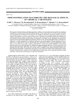
Fig. 1. Radial immunodiffusion of amphibian antibodies against sheep serum results in the development of some, typical for amphibian IgM precipitin dots and arches. C -control,
Для дальнейшего прочтения статьи необходимо приобрести полный текст. Статьи высылаются в формате PDF на указанную при оплате почту. Время доставки составляет менее 10 минут. Стоимость одной статьи — 150 рублей.