ГЕНЕТИКА, 2008, том 44, № 8, с. 1145-1147
КРАТКИЕ СООБЩЕНИЯ
УДК 575.17
Isolation of dinueleotide mierosatellite markers in commercially threatened Malaysian Mahseer, Tor tambroides © 2008 r. S. Bhassu1 , Y. Bakar2, Z. A. Rashid1
1Freshwater Fisheries Research Center, Glami Lemi, Jelebu, Negeri Sembilan, 71650 Malaysia
e-mail: subhabhassu@yahoo.com 2Faculty of Science and Technology, University Kebangsaan, Malaysia Received March 23, 2007
Seven single locus dinucleotide microsatellite markers were developed to characterize an economically important sport fish and food fish in Malaysia and in Southeast Asia. They were obtained by using a rapid method namely the 5' anchored PCR enrichment protocol. The specific primers were designed to flank the repeat sequences and these were subsequently used to characterize 120 unrelated fish from Malaysia and 30 fishes from Indonesia. The number of alleles per locus ranged from 2 (SYKVJ1-11) to 6 (SYKVJ1-4) while the levels of heterozygosity ranged from 0.0472 (SYKVJ1-11) to 0.7745 (SYKVJ1-2).
Microsatellites or simple sequence repeats (SSRs), represent a unique type of tandemly repeated genomic sequences, which are abundantly distributed across the genomes and demonstrate high levels of allele polymorphism. They are codominant markers of relatively small size, which can be easily amplified with poly-merase chain reaction. These features provide the foundation for their successful application a wide range of fundamental and applied fields of biology and medicine, including forensics, molecular epidemiology, parasitolo-gy, population genetics and genetic mapping [1, 2].
The majority of microsatellites (30-67%) found are dinucleotides. In the genome of vertebrates, (AC)„ is the most common dinucleotide motif. It is 2.3-fold nore frequent that (AT)„, the second most general type of nu-cleotides [3].
In this paper, we highlight the development of seven single locus dinucleotide microsatellite markers for Tor tambroides, based on the use of a protocol involving anchored PCR primers [4, 5].
Genomic DNA was extracted from muscle tissues using the protocol described by Qiagen Kit Protocol. One degenerate primer were used to amplify the Tor tambroides genome. The primer sequences were VJ1 (5' NNNNNNNKKVRVRV(CT)10 3'), where N = A/G/C/T; K = G/T; R = A/G; V = A/C/G; Y = T/C; H = A/C/T. A PCR reaction was carried out in a total reaction volume of 10 ^l containing ~50 ng of template DNA, 1.5 mM MgCl2, 10 mM Tris-HCl (pH 8.3), 50 mM KCl, 3 units Taq DNA polymerase (Promega, USA), 50 pmol degenerate primer respectively, 0.2 mM dNTPs (Promega, USA) and ddH2O. The PCR amplifications were performed under the following conditions: 96°C (3 min), 40 cycles of 96°C (30 s), appropriate annealing temperature (57°C for VJ1 for 20 s), 72°C (20 s) and final extension of 72°C (5 min). The PCR
The text was submitted by the authors in English.
products were then cloned into the TOPO TA cloning vector (Invitrogen, USA). Twenty recombinant clones were randomly selected for plasmid extraction, followed by DNA sequencing using the BigDye Terminator Cycle Sequencing Ready Reaction Kit (Applied Biosystems, United States) on the ABI PRISM 377 DNA Sequencer.
The primers were then designed to amplify regions containing the trinucleotide repeats using the PRIMER3 software [6], with the product length of approximately 100-300 bp. These primers were then tested for polymorphisms on 150 samples of the Malaysian Mahseer, Tor tambroides, collected from five locations in Malaysia. The PCR amplifications were performed in a 10 ^l final reaction volume containing 30 ng of genomic DNA, 10 mM Tris-HCl, 50 mM KCl, 1 unit of Taq DNA polymerase (Promega, United States), the appropriate concentration of MgCl2 and specific microsatellite primers (Table), 0.2 mM of each dNTPs (Promega, United States) and ddH2O. The amplifications were done using the Thermal Cycler (Corbett Research). The PCR conditions were as follows: 94°C (3 min) predenaturation, 35 cycles of 94°C (30 s) dena-turation, optimum annealing temperature (30 s; as shown in Table), 72°C (30 s) extension, followed by the final extension at 72°C (5 min).
The amplificons were run on a 4% Metaphor gel (BMA, United States) using 1x TBE buffer and visualized under UV light after ethidium bromide staining. The results were further validated by running on 6% non-denaturing polyacrylamide gel using 1x TBE buffer. The population data were analyzed using the POPGENE (Ver. 1.32) software [7].
The sequencing results revealed a total of 17 dinucleotide microsatellite sequences. The length of the microsatellite repeats ranged from 8 bp to 106 bp. Specific primers were then designed for 8 microsatellite loci. 1 loci were found to be monomorphic. The number of al-
1146 BHASSU Ë ap.
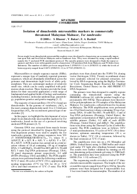
Characterization of dinucleotide microsatellite loci in Tor. tambroides
Locus Repeat motif Primer sequence (5'-3') Primer ID Exp. allele sizes, bp MgCl2, mM T * a °C Na Ho (He) GenBank accession
SYKVJ1-1 (CA)8 F: CCATGCTGTCCAGACCAATA R: GAGTGGTATCCTGCGCTCTC SYK1 214 2.5 57 3 0.3864 (0.6446) EF472538
SYKVJ1-1 (CA)8 F:TACAGTGTGGTGTGGCATCA R: GTTGGCTCTCCGTTCTTCAG SYK2 164 2.5 60 2 0.2427 (0.5436) EF472538
SYKVJ1-7 (TC)6 (TCC)3 F: CACACACACACCCACCAGAT R: TACATGGAACCACCGCTGTA SYK5 244 2.5 2.5 2.5 59 5 0.5541 (0.6806) EF472544
SYKVJ1-11 (TG)5 F: CCTCAAACGATTCCCTCAGA R: CAGCTTCTCTGTTCCAACCA SYK6 232 58 2 0.0472 (0.4873) EF472548
SYKVJ1-4 (AC)4 (CT)4 F: TCATGCTCACACACAGCACTC R: CCGGTAGCGCTCTCTGTTG SYK7 219 2.5 58 6 0.5429 (0.7672) EF472541
SYKVJ1-2 (CA)6 AA(CA)12 CC(CA)3 F: TGACCCCGTGATTTAATTCC R: AGCCATGAAAGCCATGAATC SYK8 216 2.5 58 4 0.7745 (0.7297) EF472539
SYKVJ1-16 (TG)8 F: GAGTGGTATCCTGCGCTCTC R: CCATGCTGTCCAGAGCAATA SYK9 214 2.5 60 2 0.4476 (0.5644) EF472553
Note: Ta, annealing temperature; Na, number of allele; Ho, observed heterozygosity; He, expected heterozygosity.
leles detected at each locus ranged from 2 (SYKVJ1-11) to 6 (SYKVJ1-4), while the observed heterozygosities ranged from 0.0472 (SYKVJ1-11) to 0.7745 (SYKVJ1-2) (Table).
The single locus dinucleotide microsatellite markers developed in this study are suitable for detecting polymorphisms in the Mahseer. Dinucleotides are mostly clustered in noncoding regions. In vertebrates, they are distributed 42 and 30-fold less frequently in exons than in intronic sequences and intergenic regions, respectively [3]. Nevertheless, the dinucleotides are type II markers which are very helpful for building a dense linkage map framework into which type I (coding) markers can then be incorporated. There fore, enrichment of linkage map with both set of markers will serve as important criteria in characterization of genes re-
sponsible for medically, agriculturally and evolutionary important complex traits.
ACKNOWLEDGMENTS
We would like to thank the Director of Research, E. Ismail Awang Kechik for full support in this study. This work was supported by Malaysian Fisheries Department (Inland Fisheries), grant 22501-002 (1.3).
REFERENCES
Bhassu, S.,Yusoff, K., Panandam, J.M., et al., The Genetic Structure of Oreochromis spp. (Tiapia) populations in Malaysia as revealed by microsatellite DNA analysis, Biochem. Genet, 2004, vol. 42, nos 7/8.
ÉEHETHKA TOM 44 < 8 2008
ISOIATION OF DINUCLEOTIDE MICROSATELLITE MARKERS
1147
2. Sakamoto, T., Danzmann, R.G., Gharbi, K., et al., A microsatellite linkage map of rainbrow trout (Oncorhyn-chus mykiss) characterized by large sex diffrences in recombination rates, Genetics, 2000, vol. 155, pp. 1331— 1345.
3. Toth, G., Gaspari, Z., Jurka, J., Microsatellites in different eukaryotic genomes: survey and analysis, Genome Res., 2000, vol. 10, pp. 967-981.
4. Fisher, P.J., Gardner, R.C., Richardson, T.E., Single, locus microsatellites isolated using 5' anchored PCR, Nucl. Acid Res., 1996, vol. 24, pp. 4369-4371.
5. Kumar, S.V., Tan, S.G., Quah, S.C., Yusoff, K., Isolation of microsatellite markers in mungbean, Vigna radi-ata, Mol. Ecol. Notes, 2002, vol. 2, pp. 96-98.
6. Rozen, S., Skaletsky, H.J., Primer 3. Code available at http://www.genome.wi.mit.edu/genome_software/other/ primer3.html. 1997.
7. Yeh, F.C., Boyle, T.J.B., Population genetic analysis of co-dominant and dominant markers and quantitative traits, Belgium J. Bot, 1997, vol. 129, p. 157.
ИЗОЛЯЦИЯ ДИНУКЛЕОТИДНЫХ МИКРОСАТЕЛЛИТНЫХ МАРКЕРОВ ЭКОНОМИЧЕСКИ ЦЕННОГО МАЛАЙЗИЙСКОГО ВИДА БАРБУСА Tor tambroides
© 2008 г. С. Бхассу1, Ю. Бакар2, 3. А. Рашид1
1 Freshwater Fisheries Research Center, Glami Lemi, Jelebu, Negeri Sembilan, 71650 Malaysia
e-mail: subhabhassu@yahoo.com 2 Faculty of Science and Technology, University Kebangsaan, Malaysia
Определены семь монолокусных динуклеотидных микросателлитных маркера для исследования изменчивости экономически ценного вида рыб Малайзии и юго-восточной Азии Tor tambroides. Специфические праймеры, фланкирующие повторяющиеся последовательности, были использованы для характеристики 120 особей Tor tambroides из Малайзии и 30 особей из Индонезии. Число аллелей на локус варьировало от двух (SYKVJ1-11) до шести (SYKVJ1-4), а уровень гетерозиготности -от 0.0472 (SYKVJ1-11) до 0.07745 (SYKVJ1-2).
ГЕНЕТИКА том 44 < 8 2008
Для дальнейшего прочтения статьи необходимо приобрести полный текст. Статьи высылаются в формате PDF на указанную при оплате почту. Время доставки составляет менее 10 минут. Стоимость одной статьи — 150 рублей.