ИЗВЕСТИЯ РАН. СЕРИЯ ФИЗИЧЕСКАЯ, 2015, том 79, № 1, с. 11-15
УДК 544.173.3;543.51
MASS LOSS BY SU-8 POLYMER UNDER X-RAY IRRADIATION © 2015 г. V. Nazmov1, A. Volker2, T. Fischbock2, J. Rothe3, E. Huttel2
E-mail: vladimir.nazmov@kit.edu
The principal volatile products forming in air degradation of SU-8 polymer under influence of synchrotron radiation in the wavelength range of 2.5—0.25 A have been studied. The material degradation products such as hydrogen, acrolein, acetone, allyl alcohol, benzene, and methyl formate have been identified, as well as atmospheric products. However, the most often products of outgassing are water and carbon oxide molecules, which contribute to the mass loss of the polymer irradiated.
DOI: 10.7868/S0367676515010238
1. INTRODUCTION
In the last decade, photoresist SU-8, which is based on cured diglycidyl ether of bisphenol-A (DGE-BA), has been widely used in basic investigations and industrial manufacturing. Its cross-linked structure, exhibiting a three-dimensional molecular network, allows SU-8 application as a thermosetting resin with favorable material properties, such as low shrinkage, high strength, good chemical resistance, and high electrical insulation [1, 2]. Due to its excellent stability against ionizing radiation [3], namely X-rays, cured epoxy resin has been employed for manufacturing of magnets and insertion devices at synchrotron sources, and later, due to its photosensitivity, SU-8 photoresist has been applied to manufacturing of X-ray masks [4], X-ray lenses [5], and X-ray gratings, which are used in the Talbot interferometer [6]. Nevertheless, SU-8 specimens exposed to X-rays generate volatile fragments leaving the polymer and causing slow degradation of the material [3]. This degradation results in geometrical distortions of the patterned microstructures and deterioration of their physical properties [4, 7, 8].
In this study, actual degradation conditions are reproduced, i.e. the aforementioned X-ray polymer optics are used in air atmosphere under both high X-ray power and high X-ray dose deposited, for mass-spec-trometry, which is often applied to identification of volatile degradation products [7—12].
2. EXPERIMENTAL
2.1. Materials
The SU-8 specimens of 4 inches in diameter were prepared by casting of the SU-8(10) photoresist (produced by MicroChemCorp) onto a Kapton foil and
1 Institute of Microstructure Technology, Karlsruhe Institute of Technology, Karlsruhe, 76021 Germany.
2 Institute for Photon Science and Synchrotron Radiation, Karlsruhe Institute of Technology, Karlsruhe, 76021 Germany.
3 Institute for Nuclear Waste Disposal, Karlsruhe Institute of Technology, Karlsruhe, 76021 Germany.
dried at 95°C for several hours until the residual solvent (y-butyrolactone) concentration achieved ap-prox. 3.5 weight percent. Then, a scanning X-ray beam irradiated the dried SU-8 layer through an improvised X-ray mask at the X-ray lithography station LITHO-III at the ANKA synchrotron source. The X-ray mask consists of several copper wires of 1 mm in diameter, which are non-transparent to the radiation and separated from each other by a space of 12 mm, making slit-like openings. With the spectrum of the incoming X-ray beam, set by the varying number of aluminium foils, the dose deposited was 44 and 40 J/cm3 at the top and at the bottom of the resist layer, respectively. After the post-exposure baking at 95°C for 10 hours and development with 1-Methoxy-2-propyl acetate (PGMEA), the Kapton foil was removed, releasing free standing SU-8 foils of 12 mm in width and several centimeters in length.
The degradation was analysed using two different techniques described below.
2.2. Mass Spectra Analysis
The mass spectrometry technique was employed at the TOPO-TOMO beamline (ANKA synchrotron source), where white X-ray beam was delivered to the measurement point from a bending magnet in 30 m from it.
A stack of free standing SU-8 foils of different thickness from 50 ^m to 490 ^m was placed into a measurement cell with two windows on the front and the back sides, through which the X-ray beam crossed the cell. The X-ray beam dimensions on the specimen are 25 mm (hor.) x 15 mm (vert.).
To avoid X-ray power attenuation, which might result in heating of the specimens, we inserted a stack of aluminium foils into the beam. Nevertheless, the temperature of the specimens as calculated in a hypothesis of heat losses via conduction, convection in the gas phase, and radiation [13, 14] was in the range between 65 and 92°C.
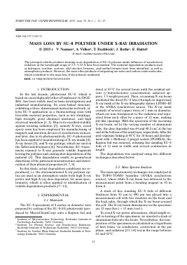
W, cm-3 • Â-1 • mA-1 8
7
6
5
4
3
2
1
No filter ......Al-foils, 51 цт Al-foils, 96 цт
/ / s // v /'•"•. ч //.' ■ \ г- * 4 г." 4 к * —г 1.1. \ 1 \ \ \ ч \ \ \ V \ 1 , " * ■ J - " "--1
0.5
1.0
1.5
2.0 2.5 Wavelength, Â
Fig. 1. Spectral distribution of X-ray beam power absorbed in the surfaces of free standing SU-8 foils at an electron energy of 2.5 GeV and an electron current of150 mA, including a 120 cm long path of transmission in air.
V2
V
V1
vJ-QH^hih
ü
Valve1
X-ray beam
QMS 200
Valve 2
Turbo pump
Membrane pump
Fig. 2. Schematic experimental setup. Vacuum meter V1 (produced by "Balzers") is based on the thermal conductivity of gases; Penning vacuum meter V2 (produced by "Pfeiffer Vacuum") employs ionization of residual gas molecules; turbo pump model 150HT Macro-Torr by Varian.
Because of the hardness of the filtered X-ray beam (Fig. 1) the difference between the doses deposited on the front and back sides of the SU-8 foils did not exceed 40%.
The cell, which was connected to vacuum meter V1 from one side and a coupling chamber (Fig. 2) from the other side, was evacuated to a residual air pressure of about 5 mbar, at which the mean free path of molecules of air did not exceed the inner diameter of the cell. The residual air in the cell played a role of a precursor gas imitating the atmospheric conditions, on the one hand, and a carrier gas delivering the volatile fragments to the mass spectrometer, on the other hand. However, the base pressure in the coupling chamber connected to the mass spectrometer was as high as of the order of 10-7 mbar, because of the direct connection to the vacuum pump.
The gas analysis was executed using a commercial quadrupole mass spectrometer (model QMS 200 F2, Prisma™, Pfeiffer Vacuum) capable of measuring ion masses of 1 to 200 Da. The ionizing chamber of the mass spectrometer was placed in approx. 140 cm from the specimens.
First of all, the cell was irradiated through the side-wall windows without specimens what resulted in out-gassing of the cell and allowed us study the volatile fragments inside as the background spectrum for further measurements. In this case, the gases typical of the air atmosphere, like H+ ions, H2O, CO, CO2, N2, and NO+ were recorded. Additionally, the mass spectra of PGMEA and isopropyl alcohol irradiated with X-rays were recorded, because these substances, when used in manufacturing of the specimens, can add some
fragments to the spectrum of the polymer during their degradation in the X-ray beam.
The interpretation of the arising spectra was carried out based on the following information: the data base with distribution of fragments of certain gases, the characteristic spectra library, the distribution of isotopes, the nitrogen rule, and the probability data for the specific ionization, and multiple ionization for a fixed filament bias [15—17].
The aforementioned data are applicable because of no any charged volatile fragments generated by direct irradiation, reach the detector of mass spectrometer, causing absence of contribution to the observed intensities. We checked this fact, switching off the filament bias. On the other hand, the recharging process and the deposition of charged fragments onto the sidewall of the transport pipe, taking into account the high collision rate under the rough vacuum condition, can suppress the flow of the charged fragments on their way toward the mass spectrometer [18].
2.3. Mass Loss Analysis
Firstly, the free-standing foils, which had been held in the evacuated cell for 12 h, were weighed on a microbalance (Mettler MT5) with an accuracy of 5 ^g. A mass loss of about 1.2% of the initial mass, ranging from 30 mg up to 350 mg, was found and ascribed to the water and y-butyrolactone evaporation from the specimens. After that, the free-standing foils were exposed to radiation and weighed periodically during the experiment.
MASS LOSS BY SU-8 POLYMER UNDER X-RAY IRRADIATION
13
Ion current, A
0 10 20 30 40 50 60 70 80 90
M, Da
Fig. 3. Ion current depending on fragment masses released from SU-8 foils irradiated by white X-ray beam from the ANKA storage ring.
3. RESULTS AND DISCUSSION
3.1. Identification of Fragments
During the irradiation with X-rays, volatile products in the mass range between 1 Da and 89 Da are observed. A corresponding mass spectrum is shown in Fig. 3, but no mass peaks in the range between 90 Da and 200 Da appeared.
In general, the volatile fragments registered are products of direct interaction with the X-rays and electrons emitted by the filament under a voltage of 90 V But, the charged fragments produced due to the electron impact only reach the detector. Mathematically, the mass spectra observed are a convolution of the mass spectrum of the volatile products stimulated by X-rays with the proper mass spectrum of each volatile product caused by the electron impact. Therefore, one can calculate the spectrum of the volatile fragments released by synchrotron radiation, deconvolving the resulting spectra and the proper spectra of the products to be recognized. These products, compounded of atoms of precursor molecules, were modelled with due account of the limited number of the products and the probability of their appearance [17].
Analysis of the appearing volatile fragments reveals three precursor types, namely the destroyed polymer, the residual gas, and water molecules dissolved in the polymer. The wat
Для дальнейшего прочтения статьи необходимо приобрести полный текст. Статьи высылаются в формате PDF на указанную при оплате почту. Время доставки составляет менее 10 минут. Стоимость одной статьи — 150 рублей.