ОПТИКА И СПЕКТРОСКОПИЯ, 2010, том 108, № 3, с. 420-425
= КВАНТОВАЯ ОПТИКА МЕТА-, МИКРО- И НАНОСИСТЕМ =
УДК 535.14
MODE MANIPULATION IN SYSTEM OF COUPLED MICROCAVITIES WITH WHISPERING GALLERY MODES
© 2010 Y. P. Rakovich*, M. Gerlach*, J. F. Donegan*, K. I. Rusakov** and A. A. Gladychchuk**
*School of Physics and CRANN Research Centre, Trinity College, Dublin 2, Ireland **Physics Department, Brest State Technical University, Brest, 224017Belarus
Received August 3, 2009
Abstract—In recent years the studies of electromagnetic modes in solid spherical microcavities have been of great interest both for their potential applicatons and fundamental optical properties. A system of coherently coupled microcavities may be called a "photonic molecule" and can be employed in the tight-binding device in order to manipulate photons in micrometer length scale. In this work we demonstrate the possibility of mode manipulation in systems of symmetric photonic molecules formed by placing several high-Q microspheres in contact. We observe photonic nanojets that reflect the symmetry of the photonic molecule, with 3 jets located at 120 degrees with respect to each other for the triangular photonic molecule. A benzene molecule-like structure consisting of a 7-microspheres cyclic photonic molecule shows a field emission pattern similar to the spatial distribution of the orbitals of the benzene molecule. We also present some results showing the coexistence of whisphering gallery modes and photonic nanojets in the same structure.
INTRODUCTION
In recent years the studies of electromagnetic modes in solid three-dimensional microcavities have been of great interest both for their potential applications and fundamental optical properties. Among others, dielectric transparent microspheres, which are three-dimensional spherical microcavities, provide high Q-factors and a small mode volume leading to strong optical feedback within the cavity [1]. The optical resonances, also called whispering gallery modes (WGMs) are caused by total internal reflection of the light at the surface inside the sphere. As the electromagnetic field is not fully confined within the sphere, an evanescent field is present surrounding the spherical particle, which enables unique possibilities for interaction of the optical resonances with the surrounding medium or objects in close proximity to the cavity. Two or more microspheres close to each other allows optical coupling of the modes between the spheres which results in a complex rearrangement of the mode structure in the strong coupling regime similar to the electronic molecular orbitals in a chemical molecule. This leads to the notation of photonic molecules (PMs) [2, 3] for coherently coupled microspheres. The optical modes ina coupled system of microcavi-ties experience splitting in to bonding and anti-bonding modes. The so called supermodes in a photonic molecule are WGMs which extend over the whole structure of the coupled microcavities.
Photonic molecule structures consisting of three or more microcavities were studied theoretically by different groups with regards to supermodes with dramatically increased Q-factors, low laser threshold and directional emission [4—7]. For optimal results, the
structure requires a symmetric photonic molecule with size-matched microcavities and optimized gap alignment between the spheres [7, 8]. Apart from microcavities arranged in arrayes, chains of coupled mi-crospheres have also been studied for waveguiding purposes [9, 10]. In our recent studies of photonic molecules made of microspheres, we investigated optical coupling of a bi-sphere system. Recently we observed the fine structure of the coherently coupled nondegenerate modes [2]. The optical coupling results in a splitting of the azimuthal resonances. In present work, we studied the optical properties of symmetrically arranged microspheres with regards to directional narrow beam emission by means of photonic nanojets [11]. These nanojets are the result of a microlens-ing effect. They emerge on the shadow side of the surface of a dielectric microcavity. The unique feature of nanojets is a directional beam with a beam waist smaller than the diffraction limit. The beam propagates over several wavelengths without significant beam divergence. Nanojets have been observed in optically coupled chains of microspheres [12]. We observed strongly directed far field emission in triangular, 5-sphere and 7-sphere structures, which we attribute to photonic nanojets. The directional emission from the PM matches the symmetry of the structure. The 7-sphere arrangement additionally shows an extraordinary hexagonal intensity distribution within the structure. Besides the observed directional emission, we carried out spectral analysis of the scattered light from the multisphere structures. Independent of the photonic nanojets, we investigated if WGMs develop within the multisphere structures. We observed uncoupled modes as well as evidence of coherently coupled modes which are present in the PM.
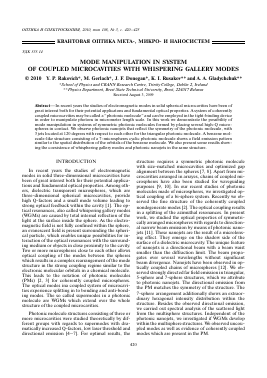
Fig. 1. Left: Photonic nanojets generated by laser illumination of a triangular photonic molecule. The far-field emission is visible due to reflection from the Si-substrate. The length scale in the frame is in micrometres. Inset top right: Image of the tri-sphere photonic molecule in white light. The crosshair indicates the focus position of the laser. Inset bottom right: A merged image of the triangular structure under laser illumination and under white light, showing the spatial distribution of the emission within the structure. Right: Image shown in inverse tones. Dark tones indicate high intensities.
EXPERIMENTAL
The PMs in different configurations were formed on a Si-wafer substrate. The microspheres used in this experiments are made of melamine formaldehyde latex (n = 1.68) with a diameter of 5.374 ± 0.069 ^m according to specifications from MicroParticles GmbH. The microspheres are coated with Rhodamine 6G fluorescence dye. This dye is used as a broadband emitter to probe the resonance spectrum of the PMs obtained by collecting the photoluminescence emission. All microspheres are touching each other so that there is no gap between the spheres. The CCD images of the emission field and the spectral measurements were taken with a commercial Renishaw micro-photolumi-nescene/micro-Raman machine. In this setup, the laser light is focused perpendicular to the substrate from above the structure through a high-NA microscope objective (100x, NA = 0.9). The scattered light is collected through the same objective operating in a 180° backscattering geometry and guided to the spectrometer and the CCD camera. The system collects all scattered light within the light cone of the objective in the focal plane. The directional emission from the PM propagates parallel to the substrate. The reflection of the beam striking the substrate is visible in the CCD images so that we can observe the directional far-field emission from the PM. The CCD images also show light distribution within the PM.
RESULTS AND DISCUSSION
The directional emission of a triangular PM is shown in Fig. 1. The laser is focused 7 ^m above the Si-substrate in the ^-direction centred in the crosshair as shown in the top inset of Fig. 1. The defocused beam allows coupling of the laser light into the whole structure, as the high-NA objective results in strong divergence of the focused beam. The position of the focus was adjusted for optimal coupling as monitored with the CCD camera. The bottom inset in the left image of Fig. 1 shows a merged image of the triangular
structure in white light and the laser illuminated image. The overlapped image shows the position where the emission developed within the triangular PM is located. As the focus position of the laser and the focus plane of the CCD camera in the micro-photoluminescence setup cannot be adjusted separately, the image of the emission is not exactly in focus. Still, the image quality is sufficient to determine details in the scattered emission. The radii of the individual spheres are about 8^. According to calculations based on Mie theory, a sub-^ nanojet on the surface of a microsphere can be obtained from spheres with a radius of about R ~ 5X and a refractive index of n ~ 1.6 [13]. Nanojets can also be formed with spheres of larger diameter (R > 20^) but with n ~ 2. With our microspheres, having a refractive index of n = 1.68, the conditions for the formation of nanojets on the spheres surface are satisfied. The image in Fig. 1 clearly shows directional emission from all three microspheres at the outer surface of the individual spheres in the structure. Most remarkable are the emission spots with high intensity located near the surface of the spheres (see bottom inset). The reflections of the nanojets on the substrate clearly demonstrate the direction of the propagating beams. The inverted tone image in Fig. 1 on the right hand side also brings out features of the internal intensity distribution within the PM. The scattered light pattern within the PM resembles the typical distribution of the electromagnetic field for a nanojet in a microsphere which forms successive circular-shaped intensity minima and maxima. The observed intensity pattern shows similarities to field calculations carried out in [13, 14]. It was shown in these papers that nanojets can be obtaind with plane wave illumination of mi-crospheres.
Our experiments demonstrate that directional emission in the horizontal plane in three well defined directions with the triangular-sphere structure is possible under the condition of perpendicular illumination of the PM with a defocused laser beam. The pho-
422
RAKOVICH et al.
-20-15 -10 -5 0 5 10 15 20
15 10
i 0
-5 -10 -15
-15 -10 -5
5 -0 -
-5 -10 -15
Fig. 2. Left: Triangular photonic molecule with attached single sphere. Light is coupled into the attached sphere by nanojet coupling. Inset top right: Image of
Для дальнейшего прочтения статьи необходимо приобрести полный текст. Статьи высылаются в формате PDF на указанную при оплате почту. Время доставки составляет менее 10 минут. Стоимость одной статьи — 150 рублей.