БИОЛОГИЧЕСКИЕ МЕМБРАНЫ, 2007, том 24, № 1, с. 105-107
УДК 577352
ONE REASON FOR INCREASED SEIZURE SUSCEPTIBILITY OF HIPPOCAMPUS COMPARED WITH NEOCORTEX
© 2007 r. Yu. I. Zilberter
Institut de Neurobiologie de la Méditerranée (INMED), INSERM U29, Parc Scientifique de Luminy, 13273 Marseille Cedex 09, France; e-mail: zilberter@inmed.univ-mrs.fr
Received 07.07.2006
In this study we compared the resting membrane potential and action potential (AP) activation thresholds of neocortical layer 2/3 and CA1 hippocampal pyramidal cells in brain slices from 6-8 day old mice. The activation threshold was -37 ± 2 mV in the neocortical pyramids (5 cells), and -50 ± 1 mV in the CA1 ones (5 cells). The observed difference in the AP activation thresholds may account for a higher excitability of hippocampus as compared to neocortex.
More than 50% of all partial epilepsies originate from foci in temporal lobe structures [1]. The high incidence of temporal lobe foci may occur because of the low seizure thresholds found in many temporal lobe structures, especially in the limbic structures of the mesial temporal lobe. In rats, the seizure threshold of the dorsal cornu ammonis (CA1) is the lowest in the brain, whereas the motor cortex has a threshold that is fivefold to sevenfold higher [2].
Pyramidal cells constitute the majority of neuronal population in both hippocampus and neocortex, therefore these cells define the level of excitability in both structures. In this study, we compared the resting membrane potential (RP) and action potential (AP) activation thresholds of neocortical layer 2/3 (L2/3) and CA1 hippocampal pyramidal cells in brain slices from 6-8 day old mice. Our results suggest that a significant difference in the AP activation thresholds may be one reason for a higher seizure threshold in neocortex compared with hippocampus.
EXPERIMENTAL
Electrophysiology. Brain slices were prepared from postnatal days (P) P6-P8 Swiss mice of both sexes. Sagittal slices (300-400 ^m), including both the neocortex and hippocampal formations, were cut using a Microm tissue slicer with the ice-cold oxygenated solution containing (in mM): 0.5 CaCl2, 7 MgSO4, and in which NaCl was replaced by an equimolar concentration of choline chloride. Slices were then transferred into oxygenated normal ACSF (Artificial Cerebro Spinal Fluid) containing (in mM): 126 NaCl, 3.5 KCl, 1.2 NaH2PO4, 26 NaHCO3, 1.3 MgCl2, 2.0 CaCl2, and 10 D-glucose, pH 7.4 at room temperature (20-22°C) for at least 1 h before use. For recordings, slices were placed into a con-
ventional fully submerged chamber superfused with ACSF (36-37°C) at a rate of 2-3 ml/min.
Patch-clamp recordings were performed using dual EPC-9 amplifiers ("HEKA Elektronik", Germany). The patch pipette solution for the whole cell recordings contained (in mM): 115 potassium gluconate, 20 KCl, 4 ATP-Mg, 10 Na-phosphocreatine, 0.3 GTP-Na, 10 HEPES, and biocytin 0.5%; pH 7.3 adjusted by KOH.
For recordings of action potentials, we used the same patch pipette solution as described for the whole cell recordings. To noninvasively estimate the cell RP we measured in the cell-attached mode the I-V relation of NMDAR channels using the patch pipette solution (in mM): NaCl 140; KCl 2.5; CaCl2 2; HEPES 10; EDTA 1; 4-AP 4; NMDA 0.01; glycine 0.01; pH adjusted to 7.3 by NaOH.
Extracellular field potentials were recorded using electrodes made from borosilicate glass capillaries of 1.2 mm OD and 0.94 mm ID (GC120TF-10, "Clark Electromedical Instruments", UK) filled with ACSF. Electrodes were positioned in L2/3 of neocortex or in the pyramidal cell layer of the CA3 subfield, and signals were amplified using DAM8A ("World Precision Instruments", UK; low filter: 0.1 Hz; high filter: 3 kHz).
Group measures are expressed as means ±SE; error bars also indicate SE.
Measurements of the action potential activation threshold. Action potentials (APs) were induced in neurons by synaptic activity and recorded at 50 kHz. APs were recorded during spontaneous seizure events initiated in the Mg2+-free solution. The first AP at each seizure was selected by hand with the following software analysis. Detection of the action potential activation threshold was performed semi-automatically off-line using the custom made software (IGOR Pro, "WaveMetrics",
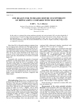
A Neocortex L2/3 pyramidal 3 min after 4-AP
Hippocampal CA1 pyramidal
200 pA
field CA1
1 min 200 |V
C
S4
rren
a2
L2/3 pyramid
JJiilUU
^.Jl Mitral „'W —20
¿[Ulouu^^
Vv^nrWv'vyvJ^ -120
_15 pA
-20 ms
8r
CA1 pyramid
-120 -80
-40 0 40 Voltage, mV
-120 -80 -40 0 40 Voltage, mV
—1—
±
s s
id id
mi mi
a a
r r
y y
p p
/3 1
2/ A
L C
o as
W S o
u
o ^
S ^
Ö n ä S w
Ö
tn
Ä a
H
o
bo o o
B
L2/3 pyramidal
100 |V 100 pA
field L2/3
5 s
w
50 | V
D
-20
-30
V m
^ -40
o
-50
P
A
-60
-70
Spontaneous synaptic activity
L2/3 pyramidal o CA1 pyramidal
0.5
1.0 1.5 2.0
Average potential slope, mV/ms
I r
B
E R T E R
A - Simultaneous recordings in L2/3 of neocortex and hippocampal CA1 region. Activities of pyramidal cells were recorded in the cell-attached mode. B - A fragment of recording in L2/3 during seizure activity. C - RP of CA1 hippocampal pyramidal cells and neocortical L2/3 ones is not significantly different. RPs were estimated by measuring the reversal potential for NMDA-activated single channel currents. D - The AP activation threshold is significantly different in L2/3 and CA1 pyramidal cells.
0
ONE REASON FOR INCREASED SEIZURE SUSCEPTIBILITY
107
USA). Only the first AP in each sequence was analyzed. The activation threshold was detected by measuring the maximum of a second derivative of membrane potential. The average potential slope was measured by averaging the first derivative of membrane potential in the time range starting from the set deviation from RP and up to the AP activation threshold.
Since the input resistance of neurons was large (522 ± ± 159 MOhm (mean ± SD) for L2/3 pyramids; 422 ± 201 MOhm for CA1 pyramids) it was necessary to correct the measured potentials by a formula:
V = V*Rseal/(Rseal - Rjn), where V* is the measured potential; Rseal is the seal resistance; Rin is the input resistance.
RESULTS
Seizures were initiated by a small concentration (50 ^M) of 4-aminopyridine (4-AP). At such concentration, 4-AP intensifies synaptic activity in the whole neuronal network without affecting any specific neurotrans-mitter receptors. Figure A shows simultaneous cell-attached recordings from L2/3 (prefrontal cortex) and CA1 pyramids in combination with field recordings (field in L2/3 not shown). The hippocampal pyramid started firing intensively shortly after application of 4-AP, while the L2/3 pyramid remained silent throughout the record. The inset (dashed line) demonstrates that firing of the CA1 pyramid is synchronous with the very initial seizure events indicating that hippocampal pyramidal cells are largely involved in the process of seizure generation. In contrast, L2/3 pyramidal cells were mostly silent during the phase of seizure initiation (Figure B).
Among the intrinsic electrical properties defining the neuron excitability are the RP and AP activation threshold. Firstly, we noninvasively measured the RP of neurons by recording the current-votage relation of NMDA-activated channels in the cell-attached mode (Figure C). In both brain regions, pyramidal cell RPs were quite hyperpolarized (see Figure C, right) and did not significantly differ from each other (p > 0.1). Thus, the level of RP is not the reason for the diverse excitability of L2/3 and CA1 pyramids.
Secondly, we tested the AP activation thresholds of both neuron types inducing firing by superfusing slices with the Mg2+-free solution. In these experiments, for seizure induction the Mg2+-free solution instead of 4-AP was used to prevent modification of APs. Figure D shows dependence of the AP activation threshold (pooled data) on the average potential slope (see Experimental procedures for details). The AP activation threshold in both cell types slightly depends on the potential slope and is significantly more negative in hippocampal pyramids. The activation threshold was -37 ± 2 mV in neocortical pyramids (5 cells), and -50 ± 1 mV in CA1 ones (5 cells). Thus, possessing RP comparable to that of L2/3 pyramidal cells, CA1 pyramidal cells have significantly more negative AP activation threshold, and that may be one reason for a higher excitability of hippocampus.
REFERENCES
1. Luciano D. Partial seizures of frontal and temporal origin // Neurol. Clin. 1993. V. 11. P. 805-822.
2. Burnham W.M. Why are complex partial seizures intractable? // Adv. Exp. Med. 2002. V. 497. P. 107-110.
Возможная причина большей чувствительности гиппокампа к судорожной активности по сравнению с неокортексом
Ю. И. Зильбертер
Институт нейробиологии, Марсель, Франция, электронная почта: zilberter@inmed.univ-mrs.fr
По сравнению с неокортексом гиппокамп обладает гораздо большей готовностью к судорожной активности. "Судороги" (пачечная активность), вызванные 4-аминопиридином в срезах мозга новорожденных мышей (возраст 6-8 дней), характеризуются активностью пирамидных клеток гиппокампа, но не пирамидных клеток префронтального неокортекса (слои 2/3). При этом потенциалы покоя пирамид в неокортексе и гиппокампе, измеренные по потенциалу реверсии NMDA-активируемыx одиночных каналов, не отличались друг от друга. В то же время порог активации потенциала действия в гиппокампальных пирамидах был значительно ниже, чем в пирамидах неокортекса (-50 ± 1 и -37 ± ± 2 мВ соответственно). Таким образом, разность в порогах активации потенциала действия пирамидных клеток может быть одной из причин различия гиппокампа и неокортекса в их возбудимости при судорожной активности мозга.
БИОЛОГИЧЕСКИЕ МЕМБРАНЫ том 24 < 1 2007
Для дальнейшего прочтения статьи необходимо приобрести полный текст. Статьи высылаются в формате PDF на указанную при оплате почту. Время доставки составляет менее 10 минут. Стоимость одной статьи — 150 рублей.