ОПТИКА И СПЕКТРОСКОПИЯ, 2014, том 116, № 2, с. 340-348
ЛАЗЕРЫ И ИХ ПРИМЕНЕНИЕ
УДК 539.18
OPTICAL EMISSION STUDIES OF SULPHUR PLASMA USING LASER INDUCED BREAKDOWN SPECTROSCOPY
© 2014 г. M. Hanif* and M. Salik**
*MCS, National University of Sciences & Technology, Rawalpindi, Pakistan **Department of Physics, School of Science, Beijing Jiaotong University, Beijing, China E-mail: drhanif-mcs@nust.edu.pk Received April 3, 2013
We present the optical emission studies of sulphur (S) plasma generated by the first (1064 nm) and second (532 nm) wavelengths of a Q-switched Nd:YAG laser. The target material was placed in front of laser beam in air at atmospheric pressure. The experimentally observed line profiles of neutral sulphur have been used to extract the electron temperature (Te) using the Boltzmann plot method, whereas, the electron number density (Ne) has been determined from the Stark broadening. The electron temperature is calculated by varying distance from the target surface along the line of propagation of plasma plume and also by varying the laser irradiance. Beside we have studied the variation of number density as a function of laser irradiance as well as its variation with distance from the target surface. It is observed that electron temperature and electron number density increases as laser irradiance is increased.
DOI: 10.7868/S0030403414020093
1. INTRODUCTION
Laser induced breakdown spectroscopy (LIBS) is an analytical detection technique for solid, liquid, and gaseous samples [1] and is based on optical detection of certain atomic and molecular species by monitoring their emission signals from the laser induced plasma [2]. It is a simple technique as compared to many other types of elemental analysis techniques because of its straightforward experimental set-up. In it, one requires a pulsed laser for generating micro plasma on the target surface and the elemental analysis is carried out by studying the emission spectrum of the plasma plume [3]. The nature and dynamic of the laser induced plasma depends on different parameters such as, laser wavelength, spot size, pulse width and ambient environment etc. By using this technique, experiments can be performed either at atmospheric pressure or in the presence of some ambient gas [4]. During ablation process, laser energy is used in dissipation into the sample through heat conduction, melting and vaporization of the target material to generate plasma plume and the main advantage of this technique is that, it provides direct chemical analysis of sample without its specific preparation [5].
Sulphur (S) occurs naturally near volcanoes and its native massive deposits are in Texas and Louisiana in the States. Its main source for industry is the hydrogen sulphide from natural gas. This element remained under investigation by many researchers since long. D. Chapman and A.G. Massey [6] presented spectroscopic studies of sulphur nitride ions. A. Banister [7]
carried out spectroscopic studies on sulphur-nitrogen compounds. J.M. Bridges and W.L. Wiese [8] presented the absolute transition probabilities and Stark widths for some prominent multiplets of S I and S II determined experimentally. David B. Beach et al. [9] presented X-ray photoelectron spectroscopic study of sulphur-nitrogen-fluorine compounds. S. Vacquie et al. [10] studied the electron temperature and number density in SF6 arc plasma. Y. Tanaka et al. [11] presented numerical computation to derive particle compositions of high-pressure SF6 plasmas in which electron temperature Te was greater than gas temperature Tg. F.G. Tomasel et al. [12] studied excitation of sulfur plasmas by a capillary discharge. G.V. Bon-darenko and Yu.E. Gorbaty [13] presented in situ Raman spectroscopic study of sulfur-saturated water at 1000 bar between 200 and 500°C. N. Konjevic et al., [14] published the critical review of selected data for the period 1989 to 2000 in which experimental stark widths and shifts for spectral lines of neutral and ionized atoms (including sulphur) were presented. Walid Tawfik and Y. Mohamed [15] presented study on calibration free laser-induced breakdown spectroscopy (LIBS) identification of seawater salinity. Rodrigo S. et al. [16] studied the applicability of a low pressure sulphur hexafluoride (SF6) plasma jet for silicon etching was investigated by optical emission spectroscopy (OES) technique. Max Wilke and Kevin Klimm [17] carried out spectroscopic studies on sulphur specia-tion in synthetic and natural glasses. P.L. Anto et al. [18] presented spectroscopic investigations and com-
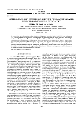
Fig. 1. Schematic diagram of experimental set up.
putational study of sulfur trioxide—pyridine complex. Jin-Tak Yeon et al., [19] presented Raman spectroscopic and X-ray diffraction studies of sulphur composite electrodes during discharge and charge.
In the present work we have employed LIBS technique for the optical emission study of the plasma generated at the surface of the target in the form of a rock stone placed in the air in front of laser beam using fundamental (1064 nm) and second (532 nm) wavelengths of a Nd:YAG laser. We report the spatial evolution of the sulphur plasma in which, experimentally observed line profiles of neutral sulphur have been used to extract the electron temperature (Te) using the Boltzmann plot method. Whereas, the electron number density (Ne) has been determined from the Stark broadening parameter. Beside we have studied the variation of electron temperature and electron number density as a function of laser irradiance.
2. EXPERIMENTAL DETAILS 2.1. The Sample
Sulphur is a multivalent non-metal, abundant, tasteless, and odorless. In its native form sulphur is a yellow crystalline solid. In nature it occurs as the pure element or as sulfide and sulfate minerals. The crystallography of sulphur is complex. Depending on the specific conditions, sulphur allotropes form several distinct crystal structures. The target material was obtained locally as rock mineral from the Balochistan province of Pakistan. It was containing mass percentage of sulphur 15.68%, iron 37.50%, molybdenum 22.41% and titanium 11.89% respectively.
2.2. The Experimental Setup
The experimental setup is shown in Fig. 1, and is same as that described in our previous work [20—25]. Briefly we used a Q-switched Nd:YAG (Quantel Bril-
liant) pulsed laser having pulse duration of 5 ns and 10 Hz repetition rate which is capable of delivering 400 mJ at 1064 nm and 200 mJ at 532 nm. The laser pulse energy was varied by the flash lamp Q-switch delay through the laser controller, and the pulse energy was measured by a Joule meter (Nova-Quantel 01507). The laser beam was focused on the target using convex lens of focal length 20 cm and diameter 30 mm. The estimated area of the laser spot on the target was ~4 x x 10-4 cm2 and 6 x 10⁻⁵ cm2 for fundamental (1064 nm) mode of Nd:YAG laser. The sample was mounted on a three dimensional sample stage, which was rotated to avoid the non-uniform pitting of the target. The distance between the focusing lens and the sample was kept at 18.5 cm, less than the focal length of the lens to prevent any breakdown of the ambient air in front of the target. The spectra were obtained by averaging 10 single data of shots under identical experimental conditions. The radiation emitted by the plasma were collected by a fiber optics (high-OH, core diameter 600 ^m) having a collimating lens (0—45° field of view) placed at right angle to the direction of the laser beam. The optical fiber was connected with the LIBS-2000 detection system (Ocean Optics Inc.), to measure the plasma emission. The emission signal was corrected by subtracting the dark signal of the detector through the LIBS software. The LIBS-2000 detection system is equipped with five spectrometers each having slit width of 5 ^m, covering the range between 220—720 nm. Each spectrometer has 2048 element linear CCD array and an optical resolution of ~0.05 nm by scanning a narrow bandwidth dye laser. In order to record the emission spectrum, the LIBS-2000 detection system was synchronized with the Q-switch of the Nd:YAG laser. The flash lamp out of the Nd:YAG laser triggered detection system through a four-channel digital delay/Pulse generator (SRS DG 535).
3. RESULTS AND DISCUSSION 3.1. Laser Induced Plasma Emission Studies
In the present work, we have used fundamental (1064 nm) and second (532 nm) wavelengths of a Q-switched Nd: YAG laser having 400 and 200 mJ pulse energy respectively, and 5 ns pulse width. The laser beam was focused on the target surface to produce the plasma by placing it in air at atmospheric pressure. In the experimental arrangement, the laser was focused on the target surface i.e., perpendicular to the target surface, whereas, the plume emission was registered as a function of the distance parallel to the surface of the target material for the spectral region from 200 to 720 nm. Fig. 2 shows the window of emission spectrum of plasma covering the spectral region from 425 to 545 nm. All the observed lines in the investigated spectral region along with their assignments are listed in Table, based on the data listed in the NBS Tables
Intensity, a.u.
3000 r
2500 2000 1500 1000
500
425 430 530 535 540 545
Wavelength, nm
Fig. 2. The emission spectrum of sulphur plasma generated by the Nd:YAG laser.
[26] and their transitions are shown in the Fig. 3. The emission spectrum was recorded covering the spectral region 300 to 600 nm as shown in Fig. 4a, whereas, Fig. 4b shows the intensity behavior of the spectrum as the distance is varied from 0.05 up to 1 mm along the direction of propagation of plasma plume generated by the second (532 nm) wavelength of the Nd:YAG laser operating at 2.5 x 10¹⁰ W cm-2.
3.2. Determination of Plasma Parameters
3.2.1. Electron temperature (Te). Determination of electron temperature is important to understand the dissociation, ionization, and excitation processes taking place
Для дальнейшего прочтения статьи необходимо приобрести полный текст. Статьи высылаются в формате PDF на указанную при оплате почту. Время доставки составляет менее 10 минут. Стоимость одной статьи — 150 рублей.