ПРИКЛАДНАЯ БИОХИМИЯ И МИКРОБИОЛОГИЯ, 2010, том 46, № 6, с. 678-684
UDC 577.154.2
PURIFICATION AND CHARACTERIZATION OF AN INTRACELLULAR P-GLUCOSIDASE FROM THE PROTOPLAST FUSANT OF Aspergillus oryzae AND Aspergillus niger © 2010 F.-M. Zhu*, B. Du**, H.-S. Gao*, C.-J. Liu***, J. Li*
*College of Food Science and Technology, Hebei Normal University of Science and Technology,
Qinhuangdao, 066600 PR China e-mail: trueyeoman@sina.com.cn, fmzhu@yahoo.com.cn **Analysis and Testing Center, Hebei Normal University of Science and Technology, Qinhuangdao, 066600 PR China ***Food Science College, Shenyang Agricultural University, Shenyang, 110161 PR China
Received May 29, 2009
Protoplasts of Aspergillus oryzae 3.481 and Aspergillus niger 3.316 were prepared using cellulose and snail enzyme with 0.6 M NaCl as osmotic stabilizer. Protoplast fusion has been performed using 35% polyethylene glycol 4.000 with 0.01 mM CaCl2. The fused protoplasts have been regenerated on regeneration medium and fusants were selected for further studies. An intracellular P-glucosidase (EC 3.2.1.21) was purified from the protoplast fusant of Aspergillus oryzae 3.481 and Aspergillus niger 3.316 and characterized. The enzyme was purified 138.85-fold by ammonium sulphate precipitation, DE-22 ion exchange and Sephadex G-150 gel filtration chromatography with a specific activity of297.14 U/mg of protein. The molecular mass of the purified enzyme was determined to be about 125 kDa by sodium dodecyl sulphate-polyacrylamide gel electrophoresis. The enzyme had an optimum pH of 5.4 and temperature of 65°C, respectively. This enzyme showed relatively high stability against pH and temperature and was stable in the pH range of 3.0-6.6. Na+, K+, Ca2+, Mg2+ and EDTA completely inhibited the enzyme activity at a concentration of 10 mM. The enzyme activity was accelerated by Fe3+. The enzyme activity was strongly inhibited by glucose, the end product of glucoside hydrolysis. The Km and Vmax values against salicin as substrate were 0.035 mM and 1.7215 prnol min-1, respectively.
p-Glucosidase, also called p-D-glucoside glucohy-drolase, catalyzes the hydrolysis of arylglucosides, alkyl-glucosides, cellobiose, and cellooligosaccharides, and is common among plants, fungi, and bacteria [1]. Many microorganisms, including fungi, bacteria, and actino-mycetes, produce enzymes that are capable of degrading plant cell walls, fulfilling an important function in the de-compostition and biogeochemical cycling of biomass. These enzymes producing from microorganisms can to be used in commercial applications and new information that allows more effective use or improvement of microbial cultures. The use of enzyme preparations as processing aids has become common practice in wine and fruit juice produciton. Winemaking is a biotechnological process in which the use of exogenous enzyme preparations helps to solve the problem of the insufficient activity of endogenous activiy in the grapes. In winemaking, flavor compounds play an important role. Monoterpenes are important constituents of the flavor of grapes, as are their fermenting products. The study of these compounds has proved that they naturally exist as nonodorous and nonvolatile glycoside forms. The aromatic potential is naturally revealed during fruit maturation by endogenous p-glucosidase. Since these enzymes have low activities and cannot liberate the whole aromatic potential, the use of
exogenous p-glucosidases in winemaking has been proposed in order to increase the amount and quality of the flavor. p-Glucosidase catalyses the hydrolysis ofcellobio-se, and to some extent cello-oligosaccharides, and therefore, completes the hydrolysis of cellulose into glucose. Fungal p-glucosidases have been isolated from various sources, such as, species of the genera Aspergillus, Botry-tis, Chaetomium, Fusarium, Humicola.
Though, there are many reports on the production of p-glucosidase from yeast (Pichia etchellsii), mesophilic fungi (Aspergillus sp.), thermophilic fungi (Melanocar-pus sp.) and hydrocarbon utilizing novel fungus (Cla-dosporium resinae) [2-5]. Recently, protoplast fusion has become a valuable technique for stain improvement. Fungal breeding using protoplast fusion techniques has been perfomed throughout the world. However, the production of the enzyme used protoplast fusion techniques has not been reported yet. This study for the first time reports the purification and characterization of p-glucosi-dases from the protoplast fusant of Aspefillus oryzae and Aspergillus niger. Some physical and chemical characteristics of the enzyme produced in media with salicin as substrate were also studied.
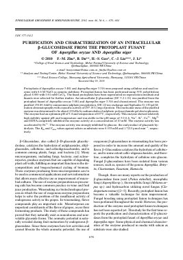
(a) (b) 0 « i 1 0° f) . * 1? 1 ,Q ® 0 (c) (d)
(e) ' "" 1HF (f) J tf (g) (h)
Fig. 1. The protoplast formation course of A. niger (a—d) and A. oryz,ae (e—h). a, e — strain native liquid; b — enzymolysis liquid after 1 h; c — enzymolysis liquid after 4 h; d, h — protoplasts after purify; f — enzymolysis liquid after 2 h; g — enzymolysis liquid after 3h.
MATERIALS AND METHODS
Materials and Chemicals. A. oryzae 3.481 and A. niger 3.316 strain were obtained from China General Microbiological Culture Collection Center. Salicin was purchased from Sigma (St. Louis, USA). DE-22, Sephadex G-150 were purchased from Amersham Pharma-cia(Sweden). SDS-PAGE Molecular weight markers for protein was purchased from Real-times (Beijing, Bio-techology Co. China). Coomassie brilliant blue was obtained from Fluka (USA) company. All other chemicals were analytical grade made in PR China.
Culture. The Czapek's medium used for the production of p-glucosidase had the composition (g l-1): sucrose - 30; NaNO2 - 2; K2HPO4 - 1; KCl - 0.5; MgSO4 - 0.5 and FeSO4 - 0.01. The pH was natural. The seed culture was prepared by inoculating a few spores from the maintenance plate into the culture medium and incubated at 27°C for 72 h. The inoculum (10%, v/v) was used for the production of enzyme in shaking flask. The shaker was run under controlled temperature (30°C), at 180 rpm for 7 days. The broth was centrifuged at 19 000 g at 4°C for 15 min and the supernatant was assayed for enzyme activity and used for purification and characterization.
Preparation of protoplasts. Protoplast was prepared from mycelia by the method previously reported [6]. The reaction mixture was centrifuged at 700 g for 10 min, and the protoplasts preciptated were suspended in a 1 ml solution (0.6 M NaCl) which was osmotically balanced.
About 100 mg (wet weight) of mycelia was added to 1 ml of enzyme solution for 2—3 h. The regeneration frequency was defined as the ratio of the number of colonies regenerating from the protoplasts and the total protoplasts used in the suspension. The real colonies regenerating from protoplasts were calculated by means of the water-lysis control test.
Protoplast fusion. The method reported previously [7—9] were used under a minor modification. Protoplasts were prepared from both biochemical mutants. Protoplasts of each mutant were mixed and then centrifuged at 700 g for 10min. This procedure was repeated several times to purify the protoplasts. The sedimented protoplasts were resuspended in 1ml of a solution of35% (w/v) polyethylene glycol (PEG) 4.000 and 0.01 mM CaCl2 • 2H2O in 0.05 M glycine-NaOH buffer (pH 7.5). After standing 25°C for 25—30 min, the suspension was centri-fuged at 700 g for 10 min. The sedimented protoplasts were resuspended in 1 ml ofan osmotically balanced medium and then plated onto hypertonic minimal medium (MM) and complete medium (CM) containing 2% agar and 0.6 M NaCl in Petri dishes, and then overlayed with the same MM containing 0.5% agar. After incubation at 25°C for 4 days, the colonies showing successful fusion were counted and the fusion frequency was estimated. The fusion frequency was defined as the percentage ratio of the number of colonies developing on MM and CM agar after the fusion treatment.
Fig. 2. The regeneration colony of A. niger (a) and A. oryzae (b, c) protoplasts.
ß-Glucosidase activity. ß-Glucosidase activities were determined in duplicate, using 0.5% (w/v) salicin as substate. Assays were performed in acetate buffer pH 4.8 and incubated at 65°C for 20 min. The amount of released glucose was determined by DNS method. ß-Glucosidase activity is expressed in units (U), defined as mol of salicin hydrolyzed by ß-glucosidase per minute, under the conditions of assay.
Protein determination. Protein concentration was determined by the Bradford method by reference to a standard calibration curve for bovine serum albumin. Protein in the column effluents was monitored by measuring ab-sorbance at 280 nm.
ß-Glucosidase extraction and purification. All purification steps were done at 4°C unless otherwise stated.The crude enzyme extract was treated with solid ammonium sulphate to obtatin the 60 to 90% fraction after centrifug-ing at 12000 g for 20 min. The precipitate was dissolved in Tris-HCl buffer (pH 7.2). Ion-exchange chromatography was performed on DE-22 in a 1.5 x 30 cm column. Proteins were eluted by stepwise NaCl concentration in-
crease, in Tris-HCl buffer (pH 7.2), with 30 ml h-1 flow rate and 3 ml fraction volume. Gel filtration was carried out on Sephadex G-150 in a 1.0 x 50 cm column, and the proteins were eluted with Tris-HCl buffer (pH 7.2), at flow rate of 12 ml h-1 and a fraction volume of 3 ml.
Electrophoresis and molecular mass estimation. Gel
electrophoresis was performed on 8% (w/v) polyacryla-mide slab gel (10 x 15 x 0.15), using a discontinuous system with 3% (w/v) stacking gel. Gels were visualized by coomassie brilliant blue staining procedure and calibrated with molecular mass markers from 43 to 200 kDa. De-naturation was achieved with 2% (w/v) SDS, heated at 100°C for 5 min.
Temperature and pH optima. Temperatures optima for activity were determined using incubations at temperatures over the range 45-80°C. Optimum pH values were determined over the pH range 3.0-6.5; purified enzym
Для дальнейшего прочтения статьи необходимо приобрести полный текст. Статьи высылаются в формате PDF на указанную при оплате почту. Время доставки составляет менее 10 минут. Стоимость одной статьи — 150 рублей.