ПРИКЛАДНАЯ БИОХИМИЯ И МИКРОБИОЛОГИЯ, 2014, том 50, № 1, с. 39-43
UDC 576.80:577.15
PURIFICATION AND CLONING OF NICOSULFURON-DEGRADING ENZYMES FROM Bacillus subtilis YB1
© 2014 Z. H. Kang*, C. C. Ren*, J. L. Zhang*, J. G. Dong**, X. Li*, and X. J. Wei*
*College of Plant Protection, Agricultural University of Hebei, Baoding 071001, Hebei Province, China; **College of Life Sciences, Agricultural University of Hebei, Baoding 071001, Hebei Province, China
e-mail: zhangjinlin@hebau.edu.cn Received February 26, 2013
The nicosulfuron-degrading enzymes from Bacillus subtilis strain YB1 were purified and their genes were cloned. The proteins of bacterial culture filtrate were precipitated with ammonium sulfate or acetone. The extracellular proteins concentrated by acetone were purified from DEAE-Sepharose Fast Flow chromatography. The four protein peaks eluted from DEAE-column were separated and purified by native PAGE. Three components (P1-1, P3-2, P4-3) had nicosulfuron-degrading activity, and component P4-3 degradated 57.5% of this compound. The molecular weights of the components were 33.5, 54.8 and 37.0 kDa, respectively. The amino acid sequences of nicosulfuron-degrading enzymes from B. subtilis YB1 were determined by MALDI-TOF-MS, indicating these enzymes as manganese ABC transporter, vegetative catalase 1 and ac-etoin dehydrogenase E1, respectively. Using PCR amplification, genes 918, 1428, 1026 bp in size were detected for the enzymes studied.
DOI: 10.7868/S0555109914010048
Nicosulfuron is the broad-spectrum sulfonylurea herbicide used in corn field at very low application rates [1] However, it has strong mobility in soil and potential risk to contaminate groundwater [1—3]. Therefore, research on the degradation of nicosulfuron in nature is important to environment protection. Microorganisms are thought to play a significant role in reducing pesticides, and many bacteria with capacity of degrading these compounds have been isolated and characterized [4—11]. Gu et al. [12] reported that 84.6% of ethametsulfuron-methyl was degraded by Pseudomonas sp. SW4 within 6 days; Xu et al. [13] isolated Pseudomonas sp. D61 and D66 and Bacillus sp. D713 from pyrazosulfuron-ethyl-contaminated soil and identified the main biodegradation products as pyrazosulfuron acid using liquid chromatography-mass spectroscopy and MS-MS techniques; Cai et al. [14] found that Methyloversatilis sp. cd-1 could degrade benazolin-ethyl from the benazolin-ethyl-wastewater treatment pool.
The earlier metabolic studies on pesticides are useful to understand that most of their biodegradation was attributed to environmental microbial degradation enzymes [15]. Preparation of degrading enzymes can give more stable and active effect than using corresponding microorganism [16, 17]. Pizzul et al. [18] reported that the ligninolytic enzymes could degrade the glyphosate, but the genes for these enzymes are unknown. The mcd gene, encoding a carbofuran hydrolase, located on a plasmid with 100-kb called pPDL11, has been cloned from Achromobacter sp. WM111 [19].
A degradative bacterium, isolated and presumptively identified as Plesiomonas sp. M6, was able to hydrolyze methyl parathion to p-nitrophenol [20]. A novel orga-nophosphate hydrolase gene designated mpd was selected from genomic library of this bacterium prepared by shotgun cloning [20]. Li et al. [21] isolated a chlo-rpyrifos-degrading bacterium Sphingomonassp. Dsp-2 and cloned the mpd gene.
A bacterium Bacillus subtilis YB1 having the capacity of degrading nicosulfuron was isolated [22]. The aim of the study was to purify the nicosulfuron-de-grading enzymes and to clone their genes from this bacterium.
MATERIALS AND METHODS
Chemicals and medium. The strain Bacillus sp. YB1 tested was stored at the laboratory of Plant Protection Institute of Pesticides of Hebei Agricultural University (China). Nicosulfuron (94.87% purity) was kindly provided by the Institute for Drug Control of Hebei Province (China). Methanol and acetonitrile were of chromatographic pure grade and other inorganic and organic chemicals were of analytical grade. The Luria Bertani (LB) solid medium was used for activating bacteria. The LB liquid medium was used for culturing of B. subtilis YB1 to produce nicosulfuron-degrading enzymes.
Obtaining culture filtrate, concentration and determination of nicosulfuron-degrading enzymes. The B. subtilis YB1 was inoculated on LB solid plate and cultivat-
ed at 30°C for 24 h, then a single colony was transferred to 500-mL Erlenmeyer flask containing 200 mL LB liquid medium and shaked at 150 rpm and 30°C for 72 h. Preparation ofextracellular filtrate containing nico-sulfuron-degrading enzymes was carried out between 0 and 4°C unless otherwise specified. The suspension was centrifuged (10000 x g for 10 min at 4°C) to remove the cell pellets. The supernatant was filtered through a 0.22-mm-pore-size membrane (Millipore, Germany). The protein of the bacterial culture filtrate was precipitated with 80% (NH4)2SO4 overnight. After the centrifugation at at 10000 x g for 30 min, the precipitate was dissolved in the smallest possible volume of 50 mM Tris-HCl buffer (pH 7.0), dialyzed against the same buffer overnight, concentrated by ultra-filtration with polyethylene glycol 20 000 and used as crude enzyme.
The protein of B. subtilis YB1 culture filtrate was precipitated with acetone too. The equivalent amount of cold acetone (—20°C) was added slowly into the culture filtrate for 2 h at 4°C. The resulting precipitate was collected, dissolved and concentrated as described above.
To detect the degradation of nicosulfuorn, the crude enzymes of B. subtilis YB1 were added to an agar medium containing 2000 mg/L nicosulfuron. The degradation spot diameter was measured for estimating the ability to degrade nicosulfuorn. Each analysis was performed in triplicate.
The concentrations of nicosulfuron in all samples were analyzed by HPLC Agilent 1200 (USA) equipped with a Zorbax XDB-C18 column (inner diameter 4.6 mm; length 150 mm). The mobile phase water— acetonitrile—acetic acid (65 : 35 : 0.1, v : v : v) with flow rate 1.0 mL/min was used. Detection of nicosulfuron was performed at 240 nm, and the injection volume was 2 ^L. Each analysis was repeated 3 times.
Enzyme purification. The concentrated B. subtilis YB1 nicosulfuron-degrading enzyme solution was loaded onto a DEAE-Sepharose Fast Flow column (1.0 x 5.0 cm) preequilibrated with 20 mM Tris-HCl,
pH 7.5 (buffer A). The column was washed with buffer A at 1.5 mL/min, and fractions containing the unbound protein were collected. A linear gradient of 0 to 1.0 M NaCl with 20 mM Tris-HCl buffer, pH 7.5 (buffer B) was then run to elute the bound material from the column. Active fractions were pooled and concentrated for further purification.
Native PAGE was performed using the modified method described by Reisfeld et al. [23]. The stacking and resolving gels contained 5.0 and 8% acrylamide, respectively. After the electrophoretic separation, the single band was isolated from the gels by sharp clean razorblade. The protein-containing gel piece was transferred to centrifugal tube containing 5 mg/L of nicosulfuron and shaked at 150 rpm and 30°C for 12 h. The gel piece without the single band was used as control. The content of nicosulfuron in the samples was determined by HPLC. All experiments were conducted in triplicate.
SDS-PAGE was performed as described by Laem-mLi [24]. The stacking and resolving gels contained 5.0% and 12% acrylamide, respectively. The molecular weights of the purified enzymes were estimated by Protein-Pak 300SW column (Waters, USA). Proteins were stained in the gel with Coomassie brilliant blue R-250. Molecular weight protein markers used for SDS-PAGE were obtained from Shanghai Sangon Biological Engineering Technology and Service Co. (China).
Proteins from the gel were isolated and sequenced in the Beijing Genomics Institute (China).
The nicosulfuron-degrading enzyme genes cloning.
Total genomic DNA was prepared from Bacillus sp. YB1 by a DNA extraction kit (Takara, China).
The nicosulfuron-degrading enzyme genes for nicosulfuron were cloned from B. subtilis YB1 using PCR-based technique. The segments of the protein sequence were obtained by MALDI-TOF-MS. The homologous proteins were searched in the protein database. The primers were designed based on the homologous proteins
(P1F, 5'-CGGGATCCATGAGACAAGGGCTGATGGC-3'; P1R, 5' -CCGCTCGAGTTATTTAAGCGCTTTAGTAA-3'; P2F, 5' - GGAATTCATGAGTTCAAATAAACTGACAAC-3'; P2R,
5'-CGGGATCCATGGCGAGAGTCATAAGCATGTC-3'; P3R, 5'-CCGCTCGAGTTAATTCAATGCCGGCTCGC-3').
PCR amplifications were conducted as follows: The 20 ^L PCR reaction mixture contained 1.75 U of Taq DNA polymerase, 10 x PCR buffer (20 mM MgCl2), 0.2 mM of each dNTP, 0.3 mM of primers and 40 ng of genomic DNA template. Amplification was performed in a T-gradient Thermal Cycler PCR (Biomatra, USA) as follows: after one cycle at 94°C for 5 min, the reaction was subjected to 30 cycles at 94°C
for 30 s, 55°C for 30 s, and 72°C for 1.2 min, with final incubation at 72°C for 10 min before cooling to 4°C.
The amplification products were recovered by DNA purification kit (Takara, China) and ligated into PMD19-T-vector (Takara, China). The ligation product was transformed into E. coli DH5a cells. The positives were selected and sequenced by Shanghai Sangon Biological Engineering Technology and Ser-
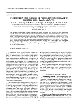
Fig. 1. Detection of nicosulfuron-degrading activity of different protein precipitates obtained from the B. subtilis YB1 culture filtrate. CK - control (50 mM Tris-HCl buffer, pH 7.0); acetone precipitation — precipitation of the enzymes with acetone; salting out — precipitation of the enzymes with (NH^SO^
vice Co. (China). The nucleotide sequences were compared with those in the GenBank database by an online alignment search.
RESULTS AND DISCUSSION
Determination of nicosulfuron-degrading enzymes precipitated by different methods. Initial exp
Для дальнейшего прочтения статьи необходимо приобрести полный текст. Статьи высылаются в формате PDF на указанную при оплате почту. Время доставки составляет менее 10 минут. Стоимость одной статьи — 150 рублей.