БИОЛОГИЧЕСКИЕ МЕМБРАНЫ, 2007, том 24, № 1, с. 43-49
== ОБЗОРЫ =
УДК 577.352
STUDIES ELUCIDATING THE MECHANISMS OF CALCIUM-INDUCED MITOCHONDRIAL DEPOLARIZATION IN INDIVIDUAL ISOLATED BRAIN MITOCHONDRIA
© 2007 r. O. Vergun
Department of Neurology, Pittsburgh Institute for Neurodegenerative Diseases, University of Pittsburgh School of Medicine, Pittsburgh, PA 15213, USA Fax: (+1) 412 648 7029; e-mail: olv1@pitt.edu Received 19.09.2006
Among other mitochondrial functions, energy production and Ca2+ uptake are crucial for maintaining neuronal viability. Both of these functions are critically dependent on mitochondrial membrane potential (A¥m). Mitochondrial Ca2+ overload causing a dissipation of A¥m is a key component of several neuronal pathologies. However, the mechanism of Ca2+-induced depolarization in neuronal mitochondria remains unclear. Typically, A¥m has been evaluated as a single overall estimate from all mitochondria present in a given cell or tissue. However, recent data showed that the population of mitochondria isolated from tissues is not homogeneous, and averaged parameters from the whole population do not necessarily reflect the processes taking place in a single organelle. This review summarizes our recent studies of Ca2+-induced depolarization in individual mitochondria isolated from rat forebrain and immobilized to coverslips. Fluorescence imaging techniques and po-tentiometric fluorescent dyes were effectively used to study A¥m changes. The data have shown that Ca2+ triggers A¥m oscillations in brain mitochondria followed by a complete depolarization. Further investigation of this phenomenon led us to suggest that Ca2+-induced A¥m oscillations can represent an intermediate unstable state that may lead to irreversible mitochondrial dysfunction. Therefore, further study of this phenomenon would help to understand what causes the irreversible damage of mitochondria during cytosolic/mitochondrial Ca2+ overload. Here we discuss the effects of different modulators of the mitochondrial permeability transition pore on Ca2+-induced depolarization in brain mitochondria and in liver mitochondria, where the mechanism of Ca2+-depolarization is better understood. A comparison of these effects in brain and liver mitochondria led us to conclude that Ca2+ can induce reversible 'low conductance' permeability transition in brain mitochondria, the phenomenon which requires a transient conformational change of the adenine nucleotide translocator from a specific transporter to a non-specific pore.
Normally functioning mitochondria establish a potential on their inner membrane (A¥m), which is typically maintained at -150 to -200 mV. A^m together with a pH gradient (ApH) is a driving force for ATP synthesis. Mitochondrial contribution to the cellular energy supply is especially important in the brain, where about 95% of total ATP is derived from oxidative phosphorylation [1]. A¥m also functions as a driving force for mitochondrial Ca2+ uptake. Driven by the electrochemical gradient, Ca2+ moves into the mitochondrial matrix through the Ca2+ uniporter located on the inner mitochondrial membrane. Mitochondrial Ca2+ uptake has a dual role in cell physiology: first, it influences ATP production via regulation of matrix dehydrogenases of the citric acid cycle; second, it regulates cellular Ca2+ signaling by buffering cytosolic Ca2+. Whereas mitochondrial Ca2+ uptake plays an important regulatory role under physiological conditions, mitochondrial Ca2+ overload is a toxic event that can lead to cell death during some pathologies.
One neuronal pathology that involves mitochondrial Ca2+ overload is excitotoxic insult, which takes place during brain hypoxia-ischemia, status epilepticus and
some neurodegenerative diseases. Under this condition, glutamate accumulated in the intracellular space over-activates glutamate receptors on the neuronal plasma membrane leading to the massive Ca2+ influx into the cytosol. The long-term activation of glutamate receptors can induce a sustained and irreversible increase of cytosolic Ca2+ concentration, which together with other factors leads to the deregulation of cellular ionic homeostasis, overproduction of free radicals, energy failure and cell death. It was shown in experiments using primary neuronal culture that a key event in the excitotoxic neuronal injury is a A¥m collapse induced by the mitochondrial Ca2+ overload [2]. Although substantial progress has been made in the understanding of the mechanisms of the neuronal death induced by excitotoxic insult [3], many questions still remain unresolved. In particular, the mechanism of Ca2+-induced depolarization of neuronal mitochondria has not been clarified. Ca2+ may dissipate A¥m by calcium cycling across the inner membrane, some form of inhibition of electron transport by the calcium overload, or mito-chondrial permeability transition pore (mPTP).
DIC
90 s
Ä
140 s
5 4 54
3 3
12 BE1B9I
180 s 645 s 690 s
Rh123 fluorescence 150 125 100 75 50 25 0
-1
-2 _l_L_
150 125 100 75 50 25
0
B
125
100
75
50
25
0
105 90 75 60 45 30 15
180 360 540 720 900 0 Time, s
1 ^m
180 360 540 720 900
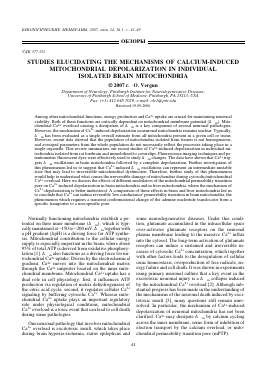
Figure 1. Examples of characteristic Ca2+-dependent changes in Rh123 fluorescence in individual brain mitochondria immobilized to the coverslip. A - Representative DIC image and fluorescent images taken at different time points illustrating A¥m oscillations. B - The plots show different patterns of spontaneous changes in Rhl23 fluorescence in five individual mitochondria from panel A. (Reprinted from [6]. Copyright 2003 by the Biophysical Society)
The studies of the mechanisms of Ca2+-induced depolarization in intact cells have limitations due to complexity of the cellular environment and a lack of direct access for drug application to mitochondria. A vast body of work studying mitochondrial physiology has been done using isolated mitochondria; this approach gives a number of advantages and usually involves experiments in a cuvette with a large population of mitochondria. However, recent data showed that the population of mitochondria isolated from tissues is not homogeneous and averaged parameters from the whole population do not necessarily reflect the processes taking place in a single organelle.
This difficulty can be avoided by using the method of fluorescence imaging of immobilized individual isolated mitochondria. This review summarizes our recent studies of the mechanisms of Ca2+-induced depolarization in individual mitochondria isolated from rat fore-brain [4-6].
MEASUREMENT OF A¥m IN INDIVIDUAL ISOLATED MITOCHONDRIA
The changes in A¥m can be studied in single isolated mitochondria immobilized to coverslip by using po-tentiometric fluorescent dyes and digital fluorescence imaging techniques. In the experiments discussed in this mini-review mitochondria were isolated from adult rat brain and liver by conventional differential centri-fugation, attached to a glass coverslip and placed into a perfusion chamber for the fluorescence microscopic measurements. The details of the method are described in [6]. The advantages of this technique compared with the cuvette experiments include (i) the ability to monitor a signal from individual organelles, as opposed to an averaged one from the whole mitochondrial population; (ii) the ability to apply and washout the drugs using a perfusion system and (iii) a negligible interaction between neighboring mitochondria due to a large volume of perfusion chamber compared with the mitochondrial volume, in particular a secondary reuptake of Ca2+ released from neighboring depolarized mitochondria which can trigger a chain reaction leading to the fast depolarization of the entire mitochondrial population in a cuvette is avoided in the experiments with attached mitochondria.
Among the fluorescent dyes suitable for measurement of A¥m, rhodamine 123 (Rh123) and tetramethylrhodamine methyl ester (TMRM) have been most frequently used to measure the changes in A¥m in experiments where the method of digital fluorescence imaging was applied. Rh123 and TMRM are lipophilic cationic dyes which are concentrated into polarized mitochondria. These two fluorophores can be effectively used to monitor changes in A¥m in individual isolated mitochondria. In our and others' experiments [6-8] Rh123 and TMRM were added to the perfusion medium in their non-quenched concentration, so that increased fluorescence signal in the mitochondrial matrix corresponded to increased A¥m, whereas diminished fluorescence indicated mitochondrial depolarization.
Ca2+-INDUCED A¥m CHANGES IN INDIVIDUAL ISOLATED MITOCHONDRIA
The measurements of A¥m in individual brain mitochondria have revealed that the population of isolated mitochondria is not homogeneous. Figure 1 demonstrates the different patterns of A¥m dynamics in the presence of very small (30-100 nM) Ca2+ concentrations. As shown, some mitochondria maintained a stable A¥m, some mitochondria depolarized irreversibly whereas the majority of mitochondria exhibited oscillations in A¥m. These A¥m dynamics were observed when mitochondria were incubated in K+-based buffer supplemented with substrates for respiratory complex I,
2 mM P O4, 5 mM Mg2+ and in the absence of adenine nucleotides. Oscillations in A¥m were previously de-
A
80 r
B
60
40
S 20
C
100
75
50
S 25
-1-1-1-1—
4 6 8 10
Ca2+
ADP
125 100 75 50 25 0
RuRed
-1-1-1-1—
8 10 12 14
D
60 45 30 15 0
ATP
CA
F 60
45
30
15
0
!
ADP
BA
-1-1-1-r
6 8 10 12 Time, min
-1-1-1-1-1—
4 6 8 10 12 Time, min
8 10 12 Time, min
Figure 2. A, B - Effect of inhibition of mitochondrial Ca2+ uptake on Ca2+-induced A¥m oscillations. C-F - Effects of mPTP modulators ADP, carboxyatractyloside (CA) and bongkrekic acid (BA) on A¥m oscillations in the isolated brain mitochondria. Each trace represents changes in A^m in individual mitochondrion. The A¥m oscillations were observed at 30-100 nM external Ca2+. (Modified from
Для дальнейшего прочтения статьи необходимо приобрести полный текст. Статьи высылаются в формате PDF на указанную при оплате почту. Время доставки составляет менее 10 минут. Стоимость одной статьи — 150 рублей.