ХИМИЯ ВЫСОКИХ ЭНЕРГИЙ, 2013, том 47, № 3, с. 226-230
НАНОСТРУКТУРИРОВАННЫЕ СИСТЕМЫ И МАТЕРИАЛЫ
SYNTHESIS AND PHOTOPHYSICS OF LEADSULPHIDE
NANOCRYSTALLITES
© 2013 г. Shipra Mital Gupta
University School of Basic and Applied Sciences, Guru Gobind Singh Indraprastha University, Dwarka Sector 16 C, New Delhi 110075 (India) E-mail: shipra.mital@gmail.com Поступила в редакцию 03.09.2012 г. В окончательном виде 29.11.2012 г.
Nanoparticles of lead sulphide have been stabilized in the presence of excess Pb2+ in aqueous basic medium by a simple chemical route of synthesis. These PbS nanoparticles were synthesized very conveniently at room temperature using hexametaphosphate as stabilizer. These nanoparticles have an absorption extending into the NIR region. A significant quantum confinement effect made the bandgap of lead sulphide nanoparticles produce a blue shift from 0.41 eV to about 1.5 eV. The size and morphology of the particles were studied by TEM. Particles were relatively small sized (about 6 nm) having narrow size distribution. XRD data analysis indicate that the product is a mixture of PbS, PbO and Pb(OH)2. Both XRD pattern and HRTEM images confirm the crystalline structure of lead sulphide crystals. IR spectroscopy indicates the formation of PbS. PbS nanoparticles were fairly stable and could be stored for about three weeks at room temperature and for about two months at 5°C without agglomeration. These particles were photoactive and sensitized the reaction of aniline by light leading to the formation of azobenzene.
DOI: 10.7868/S0023119713030058
Semiconductor nanocrystals exhibit unique electronic, optical, magnetic, photophysical and photochemical properties due to quantum size effect [1]. Transition metal chalcogenide nanoparticles are very important semiconductors because of their excellent photoelectron transformation properties and potential applications in interdisciplinary fields like solar cells, sensors, photon computers etc. [2]. Lead sulfide with a narrow gap (0.41 eV, 300 K) [3], large exciton Bohr radius (18 nm) [4] and cubic rock salt structure exhibit a strong quantum confinement effect. It has been a promising material due to its potential applications in the field of nanosized photography [5], IR detector [6], Pb2+ ion selective sensor [7], solar absorption [8] and optical switch [9, 10] and so extensive work is being carried out on it. Various methods for the preparation of PbS nanoparticles like liquid phase synthesis [11, 12], gas phase synthesis [13], polymer films route [14, 15,], synthesis in zeolites [16], block copolymer nan-oreactors [17], inverse micelle [18], microemulsion [19], gamma-irradiated non-aqueous solution [20], microwave irradiation technique [21] and epitaxial growth method [22] have been developed. New methods for the synthesis of stable lead sulfide particles in aqueous media and ambient conditions are targets pursued by material scientists. With this aim, in the present work, we have synthesized PbS nanoparticles in aqueous media and room temperature.
MATERIALS AND METHODS
All chemicals used were of analytical grade and were used without further purification — lead (II) acetate tii-hydrate, sodium hydroxide, chloroform (Merck); sodium hexametaphosphate (Fischer Scientific); hydrochloric acid (Qualigens); Ferrous sulphide (loba Chemie); aniline (Thomas Baker); N2 (analytical grade having purity >99.9%).
Electronic spectra were recorded on Carry 5000 UV-Vis-Nir spectrophotometer in the wavelength range of 200—1350 nm at room temperature. Steady-state photolysis was carried out on an Oriel photolysis assembly equipped with 200 W Hg (Xe) arc lamp. Transmission electron microscopy and selected area electron diffraction was performed by a JEOL 2100F transmission electron microscope. Samples for recording the electron micrograph were prepared by applying a small drop of the sample on a carbon coated 300 mesh copper grid and allowed to dry at room temperature before taking measurements. Electron micrographs were recorded by scanning the grid with an accelerating voltage of 200 kV at different magnifications. Solid samples prepared by removing water on DNA Speed Vic model DNA 110—230 concentrator at room temperature and 50 Hz were used for recording IR and XRD. IR spectra were recorded in far-IR range on a Thermo Scientific Model Nicolet 6700 FTIR spectrophotometer using polystyrene as media. X-ray diffraction patterns were recorded on PAN analytical X'Pert PRO diffractometer
working at 45 kV 40 mA, using Cu K radiation (k = = 0.15418 nm) in the 29 range from 25° to 60°. GC-MS data were obtained on a Thermo Fisher Scientific GC-MS, (DSQ II MS and Focus GC with Triplus Au-tosampler) MS-DSQ II and Focus Gas Chromatograph and column-TR-5MS.
The base solution of PbS was prepared at room temperature as follows: lead II acetate trihydrate solution (0.1 mol dm-3) was added drop-wise to sodium hexametaphosphate solution to obtain a reaction mixture having 3 x 10⁻⁴ mol dm-3 of each of these reac-tants. The pH of this solution was adjusted to around 10 and was flushed with nitrogen gas for about 5 min. Stoichiometric amount of SH~ was injected through a septum under vigorous stirring. The solution turned dark brown. The resulting solution was again flushed with N2 strongly for about 5 min to remove any excess SH-. 5 x 10~2 mol dm-3 of NaOH was added to establish a pH 10 followed by addition of 0.1 mol dm-3 of lead II acetate trihydrate till these contained about 6 x 10⁻⁴ mol dm-3 of excess Pb2+. The final pH of the catalyst was then adjusted to around 10.
RESULTS AND DISCUSSION
Electronic properties. The UV-Vis-NIR absorption spectrum of the PbS nanoparticles at pH 10.0 is shown in Fig. 1. Well-defined absorption features were exhibited in the UV-Vis-NIR absorption spectrum, which is due to the UV absorption of the produced PbS crystals and indicates a very narrow size. The bandgap of these lead sulphide nanoparticles is observed to be about 1.5 eV and shows a very significant blue shift from the bulk PbS crystals [17]. This is an indication of quantum confinement, showing that the average size of PbS nanoparticles is smaller than the excitonic Bohr radius of the bulk PbS (ca. 18 nm).
PbS nanparticles were most stable at 3 x 10⁻⁴ mol dm-3 sodium hexametaphosphate. At both lower as well as at higher concentrations of sodium hexametaphos-phate the stability decreased and the particles precipitated instantaneously at very low or high concentration. The lowering of pH of the reaction mixture to less than 10.0 before injecting SH~ destabilized these particles and the colloidal solution coagulated within few hours. The same lack of stability is observed at very high pH.
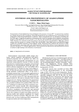
Particle size and crystalline structure. The dimensions and morphologies of the PbS nanoparticles are shown in the TEM image (Fig. 2a). From the TEM image, it is apparent that the prepared PbS nanoparti-cles are present in spherical shape. The average size of these nanoparticles is about 6 nm, which is less than the excitonic Bohr radius of the bulk PbS (ca. 18 nm). The crystal structures of these PbS nanoparticles were further studied using high-resolution TEM. Fig. 2b, 2c show the HRTEM image of PbS nanoparticles and indicates highly crystalline lattice fringes. The 0.291 nm
2.0
1.5
<D О Й св -О
3
1.0
0.5
200 400 600 800 1000 Wavelength, nm
1200
Fig. 1. UV-Vis-NIR absorption spectrum of the PbS nanoparticles at pH 10.0.
fringe spacing of the lattice was found which could be indexed as (2 0 0) of a cubic PbS crystal according to JCPDS card no. 78-1901. Apart from this, fringe spacing of 0.198 nm and 0.230 nm were also observed which corresponded to Pb(OH)2 and (2 0 0) of tetragonal PbO according to JCPDS card no. 11-0270 and 85-1739 respectively. This indicates that apart from PbS, Pb(OH)2 and PbO are also present in the sol. Fig. 2d shows selected area electron diffraction pattern (SAED) of PbS crystal, which confirms that the major of PbS prepared are in cubic crystalline phase.
The composition of the prepared nanoparticles was further analyzed using powder X-ray diffraction. The XRD pattern of the prepared nanoparticles showed the presence of broad peaks indicating the nanocrys-talline nature of the particles and confirmed the presence of cubic PbS, tetragonal PbO and Pb(OH)2 in the mixture.
Analysis of metal ion-sodium hexametaphosphate interaction. As PbS particles are associated with an excess of Pb2+, the possibility of interaction between Pb2+ and sodium hexametaphosphate was analyzed by electronic spectroscopy. The absorption spectra of blank Pb2+, sodium hexametaphosphate and their reaction mixture at pH 10.0 were recorded. The absorption of the reaction mixture was very similar to that of the sum spectra of blank Pb2+ and sodium hexameta-phosphate. This showed the absence of physical as well as chemical interaction between them as was revealed by the absence of development of any new peak in the entire recorded wavelength region (200-800 nm). The formation of PbS was further confirmed by IR spec-troscopy.
Stability. PbS was fairly stable and could be stored for about three weeks at room temperature and for about two months at 5°C without agglomeration as was revealed by the absence of any change in the shape
0
(a)
(b)
1 nm
5 A
Fig. 2. (a). TEM image, (b) & (c) HRTEM image and (d) SAED image of synthesized PbS.
2.5 r 2.0 -
o
% 1.5 -
CS
•8
o
-9 1.0 " <
0.5 -0 -
200 300 400 500 600 700 800 Wavelength, nm
Fig. 3. Absorption spectra of prepared PbS at pH 10.0 before (—) and after (■■■) heating to 80°C.
of electronic spectra. However, on keeping for longer, the color of these PbS nanoparticles faded from dark brown to light brown and showed aggregation.
Thermolysis. To further analyze the stability of these PbS particles, the effect of heating was examined by probing their electronic properties. Heating at 80°C up-to 1 hour and then cooling it to room temperature does
Для дальнейшего прочтения статьи необходимо приобрести полный текст. Статьи высылаются в формате PDF на указанную при оплате почту. Время доставки составляет менее 10 минут. Стоимость одной статьи — 150 рублей.