КООРДИНАЦИОННАЯ ХИМИЯ, 2015, том 41, № 11, с. 700-704
УДК 541.49
SYNTHESIS, CRYSTAL STRUCTURE, AND ANTIBACTERIAL ACTIVITY OF A MANGANESE(III) COMPLEX DERIVED FROM N,N'-3,4-CHLOROPHENYLENE-£w(5-METHYLSALICYLALDIMINE)
© 2015 L. Xue, D. Deng, Y. Xu, and Q. Wang*
Modern Medical Research Center, Third Affiliated Hospital of Soochow University, Changzhou, 213003 P.R. China *E-mail: wangqiangszu@126.com Received March 17, 2015
A new mononuclear manganese(III) complex has been synthesized from the Schiff base compound N,N'-3,4-chlorophenylene-bis(5-methylsalicylaldimine) and manganese perchlorate in the presence of sodium azide. The complex has been characterized by physico-chemical and spectroscopic methods, as well as single crystal X-ray determination (CIF file CCDC no. 1054335). The Mn atom in the complex is six-coordinate by two nitrogen and two oxygen atoms of the Schiff base ligand, one nitrogen atom of an azide ligand, and one oxygen atom of a methanol ligand. Crystal structure of the complex is stabilized by hydrogen bonds and %•••% interactions. The complex and the Schiff base compound were assayed for antibacterial activities against three Gram-positive bacterial strains (B. subtilis, S. aureus, and St. faecalis) and three Gram-negative bacterial strains (E. coli, P. aeruginosa, and E. cloacae) by MTT method. As a result, the complex showed effective antimicrobial activity against the microorganisms tested.
DOI: 10.7868/S0132344X15110109
INTRODUCTION
Schiff bases represent one of the most widely utilized classes of ligands in metal coordination chemistry. They offer versatile and flexible ligands capable of binding various metal ions to give complexes with versatile structures and properties [1—5]. Over the past few decades considerable study has been made on the chemistry of manganese(III) complexes derived from Schiff base ligands due to their important role in catalytic, magnetic and biological properties [6—10]. In addition, manganese plays an important role in metal-loenzymes such as catalase [11], superoxide dismutase [12, 13], and photosystem II of green plants [14, 15]. An important aspect of Mn(III) salen type complexes is their antibacterial application [16—18]. We report here the synthesis, characterization including single crystal X-ray structure of a new manganese(III) complex, [Mn(L)(N3)(MeOH)] ■ MeOH (I), where L is the dianionic form of N,N'-3,4-chlorophenylene-bis(5-methylsalicylaldimine) (H2L). The antibacterial activity against three Gram-positive bacterial strains (B. subtilis, S. aureus, and St. faecalis) and three Gram-negative bacterial strains (E. coli, P. aeruginosa, and E. cloacae) by MTT method was studied.
EXPERIMENTAL
Materials and physical methods. The Schiff base compound H2L was prepared by 2 : 1 condensation of
5-methylsalicylaldehyde and 4-chloro-o-phenylene-diamine in methanol, according to the literature method [19]. All the other reagents and solvents were purchased from commercial sources and used as received. FT-IR spectra were recorded as KBr pellets on Bruker Tensor-27. Elemental (C, H, and N) analyses were performed on a Perkin-Elmer 2400 II analyzer. Single crystal X-ray diffraction was carried out with a Bruker Apex II CCD diffratometer. Magnetic susceptibility measurement was carried out with a Sherwood Scientific Co., UK magnetic susceptibility balance. Electronic spectra were obtained with Lambda 900 spectrophotometer. Molar conductivity of the complex in acetonitrile was measured with a DDS-11A molar conductivity meter.
Caution! Perchlorate and azide complexes of metal ions are potentially explosive. Only a small amount of material should be prepared, and it should be handled with caution.
Synthesis of complex I. To a stirred suspension of H2L (0.378 g, 1.00 mmol) and sodium azide (0.130 g, 2.00 mmol) in methanol (20 mL) was added dropwise a methanol solution (10 mL) of manganese(II) perchlorate hexahydrate (0.254 g, 1.00 mmol). After a few minutes a brown precipitate started to deposit. This was dissolved by adding the requisite amount of acetonitrile. After one hour stirring, the solution was filtered and the filtrate was kept for slow evaporation. The diffraction quality deep brown single crystals that depos-
Table 1. Crystallographic data and refinement parameters for complex I
Parameter Value
Molecular weight 537.88
Crystal color; habit Brown; block
Crystal size, mm 0.18 x 0.18 x 0.13
Crystal system Monoclinic
Space group P2j/n
Unit cell dimensions:
a, A 17.185(2)
b, A 7.477(1)
c, A 19.455(3)
P, deg 101.898(2)
V, A3 2446.1(6)
Z 4
Pcalcd g cm-3 1.461
p., mm-1 0.690
9 Range collected, deg 2.42-25.50
Tmin and Tmax 0.886 and 0.916
Reflections collected/unique 21 588/4534
Observed reflections (I > 2ct(I)) 3746
Data/restraints/parameters 4534/2/334
R1, wR2 (I > 2ct(T>) 0.0375, 0.0956
R1, wR2 (all data) 0.0499, 0.1020
GOOF on F2 1.066
Largest differences in peak/hole, e/A3 0.299/-0.267
ited over a period of a few days were collected by filtration and washed with methanol. The yield was 435 mg (81%).
For C24H25N5O4ClMn
anal. calcd., %: Found, %:
C, 53.59; C, 53.45;
H, 4.68; H, 4.77;
N, 13.02. N, 13.15.
Crystal structure determination. Intensity data of complex I were collected at 298(2) K on a Bruker Apex II CCD diffractometer using graphite-monochromat-ed Mo^a radiation (X = 0.71073 A). For data processing and absorption correction the packages SAINT and SADABS [20] were used. The structure was solved by direct and Fourier methods and refined by full-matrix least-squares based on F2 using SHELXL-97 [21] package. The non-hydrogen atoms were refined aniso-tropically. The remaining hydrogen atoms have been placed at geometrical positions with fixed thermal parameters. The Cl atom is disordered over two sites and modeled accordingly, with occupancies of 0.89(1) and 0.11(1). Crystallographic data are summarized in Table 1. Selected bond lengths and angles are listed in Table 2.
Supplementary material for structure I has been deposited with the Cambridge Crystallographic Data Centre (no. 1054335; deposit@ccdc.cam.ac.uk or http:// www.ccdc.cam.ac.uk).
Antibacterial activity. Antibacterial activity of the complex was tested against B. subtilis, S. aureus, St. faecalis, P. aeruginosa, E. coli, and E. cloacae using MTT medium. The minimum inhibitory concentrations (MICs) of the complex were determined by a colorimetric method using MTT dye [22]. A stock solution of the complex (50 ^g mL-1) in DMSO was prepared and quantities of the complex were incorporated in specified quantity of sterilized liquid medium. A specified quantity of the medium containing the com-
Table 2. Selected bond distances (A) and angles (deg) for complex I
Bond d, Â Bond d, Â
Mn(1)-O(1) 1.8651(15) Mn(1)-O(2) 1.8766(15)
Mn(1)-N(1) 1.9930(17) Mn(1)-N(2) 1.9868(17)
Mn(1)-O(3) 2.4548(18) Mn(1)-N(3) 2.229(2)
Angle ro, deg Angle ro, deg
O(1)Mn(1)O(2) 90.51(7) O(1)Mn(1)N(2) 172.88(7)
O(2)Mn(1)N(2) 93.40(7) O(1)Mn(1)N(1) 93.19(7)
O(2)Mn(1)N(1) 173.53(7) N(2)Mn(1)N(1) 82.39(7)
O(1)Mn(1)N(3) 99.33(8) O(2)Mn(1)N(3) 96.36(8)
N(2)Mn(1)N(3) 86.15(8) N(1)Mn(1)N(3) 88.28(7)
O(1)Mn(1)O(3) 90.43(7) O(2)Mn(1)O(3) 88.47(7)
N(2)Mn(1)O(3) 83.73(7) N(1)Mn(1)O(3) 86.20(7)
N(3)Mn(1)O(3) 169.04(7)
702
XUE et al.
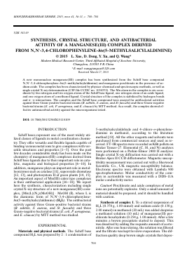
Fig. 1. Molecular structure of complex I with 30% thermal ellipsoids.
plex was poured into microtitration plates. Suspension of the microorganism was prepared to contain approximately 105 cfu mL-1 and applied to microtitration plates with serially diluted complexes in DMSO to be tested, and incubated at 37°C for 24 h for bacteria. After the MICs were visually determined on each microtitration plates, 50 ^L of phosphate buffered saline (PBS 0.01 mol L-1, pH 7.4: Na2HPO4 ■ 12H2O 2.9 g, KH2PO4 0.2 g, NaCl 8.0 g, KCl 0.2 g, distilled water 1000 mL) containing 2 mg mL-1 of MTT was added to each well. Incubation was continued at room temperature for 4-5 h. The content of each well was removed, and 100 ^L of isopropanol containing 5% 1 mol L-1 HCl was added to extract the dye. After 12 h of incubation at room temperature, the optical density (OD) was measured with a microplate reader at 570 nm.
RESULTS AND DISCUSSION
Reaction of manganese(II) perchlorate and H2L in the presence of sodium azide produces the mononuclear manganese(III) complex I. Clearly, aerial oxidation of manganese(II) to manganese(III) and metal assisted deprotonation of the phenolic moieties take place during the formation of the complex. The poor conductivity of the complex (15 fi-1 cm2 mol-1) indicates that azide ion is coordinated to the metal center and is not dissociated by acetonitrile molecule in solution. The characteristic imine stretching of the complex is observed at 1605 cm-1 as a strong signal. Appearance of intense band at 2037 cm-1 indicates the presence of azide ligand. The IR spectrum of the complex exhibits one broad and weak absorption centered at 3432 cm-1 due to the vibration of the hydroxyl groups of the methanol molecules. UV-Vis spectrum of the complex exhibits two typical bands centered at 415 and 312 nm which can be assigned to phenolate ^ ^ manganese(III) charge transfer and manga-
nese(III) ^ imine (d ^ n*) metal to ligand charge transfer transitions, respectively. The observed magnetic moment at 300 K of the complex is 4.70 B. M., indicating that the manganese(III) center in the complex exists in high spin state.
The components and crystal structure of the complex are shown in Fig. 1. The asymmetric unit of complex I contain a mononuclear [Mn(L)(N3)(MeOH)] molecule and a methanol molecule of crystallization. The structure shows that the complex having the metal center in the salen-type cavity of L2-. The Mn atom is six-coordinated, with two oxygen and two nitrogen atoms of the Schiff base ligand in the equatoiral plane, and with one azido nitrogen and one methanol oxygen atoms defining the axial positions. The coordination environment of the metal ion is
Для дальнейшего прочтения статьи необходимо приобрести полный текст. Статьи высылаются в формате PDF на указанную при оплате почту. Время доставки составляет менее 10 минут. Стоимость одной статьи — 150 рублей.