ЖУРНАЛ ФИЗИЧЕСКОЙ ХИМИИ, 2010, том 84, № 9, с. 1712-1717
ФИЗИЧЕСКАЯ ХИМИЯ НАНОКЛАСТЕРОВ И НАНОМАТЕРИАЛОВ
y%K 541.128
SYNTHESIS OF WELL-ALIGNED CARBON NANOTUBES ON THE NH3 PRETREATMENT Ni CATALYST FILMS
© 2010 Gang Li
Laboratory of Ministry of Education for Conveyance and Equipment, School of Mechanical and Electrical Engineering,
East China Jiaotong University, Nanchang, P.R. China E-mail: ligang0794@163.com Received September 05, 2009
Abstract — Well aligned multi-walled carbon nanotubes (CNTs) have been synthesized on large area Ni-de-posited SiO2/Si substrates via the pyrolysis of C2H2 using thermal chemical vapor deposition technique at 900°C. We concluded that NH3 pretreatment was very crucial to control the surface morphology of catalytic metals and thus to achieve the vertical alignment of CNTs. With higher density of the Ni particles, better alignment of the CNTs can be obtained due to steric hindrance effect between neighboring CNTs. The degree of crystallization of the CNTs enhanced with the increase of the NH3 pretreatment time was investigated by X-ray diffraction and transmission electron microscope studies. Energy dispersive X-ray spectrum analysis revealed that CNTs grew by a tip growth mechanism.
INTRODUCTION
Since the first discovery of carbon nanotubes (CNTs) by Iijima in 1991 [1], they have been promising for many potential technological applications due to their extraordinary chemical and physical properties. Synthesis of carbon nanotubes for mass production has been achieved by several methods such as laser vaporization [2], arc discharge [3, 4] and chemical vapor deposition (CVD) [5—17]. Synthesis of well-aligned CNTs on a large area is inevitably necessary for field emission device applications. Arc discharge and laser vaporization techniques can produce a large amount of CNTs, but it is very difficult to obtain uniform alignment on a large area. The CVD process has been developed for growth of CNTs in well-aligned configurations. Terrones et al. [18] fabricated vertically aligned carbon nanotubes on pulse-laser deposited Co films on silica substrates by means of thermal CVD. Li et al. [19] used a method based on CVD catalyzed by Fe nanoparticles embedded in mesoporous silica and produced large areas of vertically well-aligned long carbon nanotubes. Fan et al. [20] synthesized aligned nanobubes on Fe-patterned porous Si substrates using thermal CVD. Howerer, detailed studies of the effects of the structures of metal films on the growth of nanotubes have not been hitherto performed.
In this article, we report that very uniform, high-density nano-sized catalysts were obtained by NH3 etching pretreatment from a homogenous Ni layer. Well-aligned, vertically arranged and high density CNTs can be synthesized by thermal CVD on optimally broken catalysts. The objective of this study is to get reproducibly the catalyst nanoparticles not only with
the appropriate size and distribution but also with the high density suitable to promote the growth of densely-packed vertically aligned CNTs suitable for future application.
EXPERIMENTAL
The substrates used in this study are N-type Si (111) wafers coated with SiO2 by thermal oxidation. The SiO2 acts as a diffusion barrier between Ni and Si preventing the formation of nickel silicide. Ni catalyst films were deposited by K575X Peltier Cooled High Resolution Sputtering Coater with base pressure <10-5 mbar (Quorum/Emitech, English). The film thickness was monitored in situ by a quartz balance, and calibrated ex situ by atomic fore microscopy (AFM, Veeco Explorer).
CVD was carried out in a horizontal quartz tube (60 mm in diameter; 1200 mm in length) at atmospheric pressure. The substrates were then cut into small (20 mm x 30 mm). In each experiment, one sample was placed in the middle of the CVD tube. The quartz tube was first purged by N2 gas for 15 min to exclude air. A constant flow rate of N2 was maintained while the reaction chamber was then heated up to 600°C, which was held constant in H2 instead of N2 and Ni catalyst deoxidized for 60 min. After that, the reaction chamber was continuously heated up to the reaction temperature in hydrogen. Prior to deposition reaction, to form the nanometer-sized catalyst particles required for well-aligned CNTs growth, the Ni catalyst film was pretreated by NH3 gas, with a flow rate of 100 cm3/min for 10—60 min at a temperature
SYNTHESIS
1713
nm
-221.29
u,
^m 10
nm 120.22
5 (b)
10 ^m
10 ^m
5
0
5
0
5
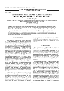
Fig. 1. AFM images for the surface morphology of the Ni catalytic nanoparticles formed on SiO2/Si substrate from 20 nm Ni film after the heat and NH3 pretreatment for at 900°C for (a) 20, and (b) 60 min.
range of 800—950°C. After the NH3 treatment, C2H2 gas was introduced into the reaction chamber at a flow of 30 cm3/min at the same temperature as the NH3 pretreatment. The growth time was varied from 5 to 20 min. Finally, the chamber was slowly cooled down to the room temperature under N2 ambient after the growth.
As-pretreated Ni catalyst films were characterized by AFM in tapping mode at ambient conditions. The morphology of as-grown CNTs were examined by field emission scanning electron microscope (FESEM, JEOL JSM-7001F). Transmission electron microscope (TEM; JEOL, JEM-2100) was used to investigate the wall structure of an individual CNT and the
crystallinity of the CNTs. To prepare the TEM samples, carbon nanotubes were peeled off by tweezers from the SiO2/Si substrates and ultrasonicated in eth-anol for 45 min to form a well-dispersed solution which was then dropped onto a 3 mm Cu grid coated with a layer of holey carbon. TEM test was performed after the grid was dried in an oven. The chemical compositional analysis of the as-deposited CNTs was carried out by energy dispersive X-ray (EDX, Oxford, Inca Energy 350) spectrum system. X-ray diffraction studies (XRD, X-Ray Diffractometer, D/max 2500 PC, CuKa radiation) were performed to identify the products and determine their degree of crystallization.
1714
GANG LI
Fig. 2. SEM morphology of the CNTs grown by thermal CVD process on the SiO^Si substrates after the NH3 etching pretreatment of (a) 20, and (b) 60 min. The insets show high magnification cross section views of (a) and (b), respectively.
Fig. 3. TEM images of the CNTs grown by thermal CVD process on the SiO2/Si substrates after the NH3 etching pretreatment of (a) 20 and (b) 60 min.
RESULTS AND DISCUSSION
AFM images of surface morphology of Ni nano-particles transported from the Ni films indicate the effect of the pretreatment conditions. Figure 1 is the AFM image in contact mode, which shows the surface morphologies of the 20 nm thickness Ni films prepared at different NH3 etching treatment conditions. In these cases, the etching treatment time were changed while fixing the flow rate of NH3 with 100 cm3/min and the etching temperature at 900°C. It is seen that the Ni films breaks into small islands and particles due to surface tension as well as the compres-sive stress resulting from the mismatch of the thermal expansion coefficients of Ni and SiO2. The Ni particle size decreases with the increase of the etching treatment time, whereas the particle density increased. Root-mean-square (RMS) roughness was measured over the whole area. According to AFM analysis we can calculate the RMS roughness of Fig. 1a and b, which are 7.449 and 3.126 nm, respectively. The NH3
etches metal film surface and decreases the surface roughness with the increasing of NH3 pretreatment time. The Ni particles are uniformly distributed over the whole surface of the substrate as shown in Fig. 1b compared to that shown in Fig. 1a, which is also confirmed by the RMS height in Fig. 1. It is concluded that the size and area density of these nanoparticles depend on the NH3 etching treatment conditions and the appropriate size distribution and high density of particles can be obtained by prolonging the NH3 pre-treatment time.
Figures 2a, b are SEM micrographs of the CNTs grown on 20 nm thick Ni films with different etching pretreatment time. The C2H2 gas for CNTs growth is fixed to the flow rate of 30 cm3/min, for 10 min at 900°C, but the NH3 pretreatment condition is changed to control the size and number density of the Ni catalytic particles. The edges are peeled off using tweezers to observe and analyze the orientation and physical properties. For the case of NH3 pretreatment with
SYNTHESIS OF WELL-ALIGNED CARBON NANOTUBES
1715
<N О О
о
(a)
О
Й
20
40
60
80
29, degree
Fig. 4. XRD of the CNTs grown on the substrates after the NH3 etched at 900°C for (a) 20 and (b) 60 min.
20 min, because of a lower particle density, the nano-tube density is also lower and the nanotubes are not strictly oriented vertically as shown in Fig. 2a. When NH3 pretreatment time prolonged up to 60 min, the nanotubes are oriented perpendicular to the surface of the substrate due to high density of Ni nanopartilces. A single nanotubes has a curly shape as shown in the insets of Fig. 2b. From Fig. 2a and b, we can also deduce that the length of CNTs increase from about 4 to 9 ^m. The insets show that the ranges of the diameter of the two kinds of CNTs decrease from 60—250 nm to 20—40 nm with increasing NH3 pretreatment time.
Since the particle size of catalyst determines the diameter of CNTs, the smaller size of catalytic particles results in a smaller diameter of CNTs. At the same time, when the density of catalytic metal particle increases, the vertical alignment degree of CNTs is significantly enhanced. When the density of CNTs reaches certain high level, the growth in non-vertical direction is prohibited due to the steric hindrance from adjacent CNTs. The vertically aligned CNTs can bundled together by van de Walls force. However when the nanoparticles density is not high enough, the lack of van der Waals interactions between neighboring CNTs results in entangled CNTs, which are observed in Fig 2a. It str
Для дальнейшего прочтения статьи необходимо приобрести полный текст. Статьи высылаются в формате PDF на указанную при оплате почту. Время доставки составляет менее 10 минут. Стоимость одной статьи — 150 рублей.