KOOPMHH^HOHHÂS XHMH3, 2015, moM 41, № 5, c. 301-305
yffK 541.49
SYNTHESIS, STRUCTURE, AND MAGNETIC PROPERTY OF A 3D SUPRAMOLECULAR Gd(III) COORDINATION POLYMER WITH 4,4-SULFONYLDIBENZOIC ACID LIGANDS
© 2015 X. N. Zhang and Z. B. Han*
College of Chemistry, Liaoning University, Shenyang, 110036 P.R. China *E-mail: ceshzb@lnu.edu.cn Received September 9, 2014
A new coordination polymer [Gd(Sfdb)(NO3)(DMF)2] • 2H2O (Sfdb = 4,4'-sulfonyldibenzoic acid) has been synthesized under hydrothermal conditions and characterized by elemental analysis, IR, TG and single-crystal X-ray diffraction (CIF file CCDC no. 1023460). The X-ray diffraction analysis shows that I (C20H24N3Oi2SGd) crystallizes in the monoclinic crystal system, which reveals that I is a 3D supramolecular framework assembled by the intermolecular hydrogen bonds. Moreover, the magnetic studies of I showed that there exist antiferromagnetic interactions between the Gd(III) centres. The unit cell parameters for I: a = = 33.453(3), b = 10.5469(10), c = 18.7895(18) Â, P = 123.7670(10)°, V = 5511.0(9) Â3, Z = 8. DOI: 10.7868/S0132344X15050096
INTRODUCTION
For decades, the design and synthesis of novel metal-organic frameworks (MOFs) have been a field of rapid growth in materials chemistry [1, 2] due to their exceptionally artistic architectures and potential applications, such as magnetic, gas adsorption and separation, heterogeneous catalysis and luminescence [3—8]. Meanwhile, crystal engineering of supramolecular polymers is also becoming popular not only due to the easily predicted H-bonding supramolecular synthons [9, 10], but also due to their potential applications as functional materials. To achieve these kinds of functional materials, it is necessary to precisely position the synthons in the crystal lattice by means of appropriate non-cova-lent interactions between molecules. Carboxylic acids amides are commonly used functional synthons in crystal engineering because they easily form robust architectures via O-H---O H-bonding [11, 12]. This approach can introduce new functions into the system of supramolecular polymers. Thus, continuing efforts have been devoted to the purpose of designing and synthesizing novel molecular architectures [13]. In this paper, we report on the synthesis, crystal structure and magnetic properties of [Gd(Sfdb)(NO3)(DMF)2] • • 2H2O (I) (Sfdb = 4,4'-sulfonyldibenzoic acid), constructed from Sfdb ligands with Gd3+ ions, which exists antiferromagnetic interactions between the Gd(III) centers.
EXPERIMENTAL
Materials and methods. All solvents and reagents employed were commercially available and used without further purification. The C, H, and N microanal-
yses were carried out on a PerkinElmer 240 elemental analyzer. Infrared spectra were recorded on the powder samples of a crystal embedded in KBr pellets from 400 to 4000 nm at a speed of 100 nm/min. The magnetic data were collected on a Quantum Design MPMS SQUID-XL-5 magnetometer using the crushed single-crystal samples. Magnetic data were corrected for the diamagnetic contribution calculated from Pascal constants [14] and a background of the sample holder.
Solvothermal synthesis of I. A reaction mixture of Gd(NO3)3 • 6H2O (0.015 g, 0.033 mmol), H2Sfdb (0.010 g, 0.033 mmol), DMF (1 mL) and C2H5OH (0.5 mL) was stirred for 20 min in air to form a solution, the solution was placed in vial (5 mL), then the vial was sealed and heated at 348 K for three days, followed by slow cooling (5 K/h). The colorless block crystals were washed with DMF and dried in air (the yield was ~62%).
For C^H^O^SGd
anal. calcd., %: Found, %:
C, 34.89; C, 34.92;
H, 3.48; H, 3.50;
S, 4.65. S, 4.66.
IR spectrum (KBr; v, cm-1): 3422 m, 2934 m, 1646 s, 1567 m, 1473 s, 1385 s, 817 w, 738 m.
X-ray crystal determination. Single-crystal X-ray diffraction measurements were collected at room temperature with a Bruker Apex II diffractometer with Mo^a radiation (X = 0.71073 Â) and graphite mono-chromator using the «-scan mode. The structure was solved by direct methods and refined on F2 by full-matrix least-squares using SHELXTL [15]. All non-hydrogen
Table 1. Crystallographic parameters and summary of data collection for structure I
Parameter Value
Formula weight Crystal system Space group
a, A
b, A
c, A P, deg V, A3 Z
Pcalcd mg cm-3 Absorption coefficient, mm-1 /(000) 9 Range, deg
Reflections collected/unique
Rint
T, K
Data/restraints/parameters Final R indices (I > 2(I))* R1 = 0.0571, wR2 = 0.1455 R indices (all data) R1 = 0.0737, wR2 = 0.1528
Largest diff. peak and hole, e/A3 1.875 and -1.982
*R1 = S||F0| - |Fc ||/S|F0|; WR2 = 2[w(^ - F2 )2]AW()2]1/2.
atoms were treated anisotropically. Positions of hydrogen atoms were generated geometrically. Crystallographic data and experimental details for structural analyses are summarized in Table 1. Selected bond lengths and angles are listed in Table 2. Hydrogen bonding geometric data is listed in Table 3. Supplementary material has been deposited with the Cambridge Crystallographic Data Centre (no. 1023460; deposit@ccdc.cam.ac.uk or http://www.ccdc.cam. ac.uk).
RESULTS AND DISCUSSION
Single-crystal X-ray diffraction analysis reveals that I exhibits a 3D supramolecular framework structure. The asymmetric unit consists of one crystallo-graphically independent Gd(III) center, one Sfdb ligand, one coordinated nitrate ion, two coordinated DMF molecules and two lattice water molecules. Each Gd(III) center is eight-coordinate and surrounded by four oxygen atoms of four different Sfdb ligands, two oxygen atoms of one nitrate ion and two oxygen atoms of two different DMF molecules (Fig. 1). As shown in Fig. 2, two Gd centres are linked by four carboxylate groups via syn-syn mode, which linked two nitrate ions and four DMF molecules to form a [Gd2(COO)4(NO3)2(DMF)4] secondary building unit (SBU). In the whole structure, Gd-O bond lengths ranging from 2.096(9) to 2.242(10) A and the OGdO bond angles are in the range of 52.0(2)° and 154.2(2)°
687.73 Monoclinic
C2/c 33.453(3) 10.5469(10) 18.7895(18) 123.7670(10) 5511.0(9) 8
1.658 2.544 2728 2.07-27.41 16960/6232 0.0454 296(2) 6232/0/329
Table 2. Selected bond lengths (A) and angles (deg) for I*
Bond d, A Bond d, A
Gd(1)-O(1) 2.314(6) Gd(1)-O(6)#3 2.355(6)
Gd(1)-O(2)#1 2.348(6) Gd(1)-O(7) 2.432(6)
Gd(1)-O(8 2.349(6) Gd(1)-O(10) 2.478(6)
Gd(1)-O(5)#2 2.350(6) Gd(1)-O(9) 2.519(6)
Angle ю, deg Angle ю, deg
O(1)Gd(1)O(2)#! 121.6(2) O(6)#3Gd(1)O(7) 77.0(2)
O(1)Gd(1)O(8) 148.7(2) O(1)Gd(1)O(10) 83.2(2)
O(2)#1Gd(1)O(8) 73.3(2) O(2)#1Gd(1)O(10) 133.9(2)
O(1)Gd(1)O(5)#2 76.2(2) O(8)Gd(1)O(10) 106.3(2)
O(2)#1Gd(1)O(5)#2 73.1(2) O(5)#2Gd(1)O(10) 77.3(2)
O(8)Gd(1)O(5)#2 134.6(2) O(6)#3Gd(1)O(10) 146.8(2)
O(1)Gd(1)O(6)#3 76.8(2) O(7)Gd(1)O(10) 73.3(2)
O(2)#1Gd(1)O(6)#3 79.3(2) O(1)Gd(1)O(9) 129.1(2)
O(8)Gd(1)O(6)#3 80.0(2) O(2)#1Gd(1)O(9) 85.2(2)
O(5)#2Gd(1)O(6)#3 122.0(2) O(8)Gd(1)O(9) 75.8(2)
O(1)Gd(1)O(7) 79.4(2) O(5)#2Gd(1)O(9) 71.8(2)
O(2)#1Gd(1)O(7) 143.2(2) O(6)#3Gd(1)O(9) 154.2(2)
O(8)Gd(1)O(7) 75.2(2) O(7)Gd(1)O(9) 104.9(2)
O(5)#2Gd(1)O(7) 143.6(2) O(10)Gd(1)O(9) 52.0(2)
* Symmetry transformations used to generate equivalent atoms: #1 -x + 1/2, -y + 3/2, -z + 1; #2 x + 1/2, -y + 3/2, z + 1/2; #3 -x, y,
-z + 1/2.
КООРДИНАЦИОННАЯ ХИМИЯ том 41 № 5 2015
SYNTHESIS, STRUCTURE, AND MAGNETIC PROPERTY
303
Table 3. Geometric parameters of hydrogen bonds in I
D H-A Distance, A Angle DHA, deg
D-H H-A D-A
C(4)-H(4^)-O(4)i 0.93 2.54 3.393(13) 152
C(7)-H(7^)-O(1) 0.93 2.44 2.753(12) 100
C(9)-H(9^)-O(4)i 0.93 2.45 3.380(11) 174
C(13)-H(13^)-O(3)u 0.93 2.36 3.236(11) 157
C(17)-H(17^)-O(8) 0.93 2.47 3.007(12) 117
* Symmetry codes: i —x, 1 — y, — z; ii —x, 2 — y, —z.
(Table 2). In compound I, Sfdb ligand adopts ^2-car-boxylato-K1O':K1O' mode, each Sfdb ligand links four Gd3+ ions. Adjacent SBUs are bridged by two Sfdb ligands to form a 1D chain (Fig. 3). And adjacent chains are further connected via the strong C—H—O hydrogen bonding interactions (Table 3) to form a 3D supermolecular framework (Fig. 4). The hydrogen bonding interactions enhance the stability of the framework.
The IR spectrum of I shows characteristic bands of carboxyl groups at 1646 cm-1 for the antisymmetric stretching and at 1385 cm-1 for symmetric stretching. The separations (A) between vas(CO2) and vs (CO2) indicate the presence of and bidentate bridging (261 cm-1) coordination modes in I [16]. The absence of strong peaks around 1720 cm-1 in I indicates that all carbox-ylic groups are deprotonated [17], which is consistent with the results of the valence sum calculations. Ac-
cording to near 3422 cm-1 strong and broad absorption peaks attributed to the O-H vibration absorption of water molecules.
The temperature-dependent magnetic susceptibilities of compound I were investigated at a temperature range of2-300 K, under an applied field of1000 G, in the form %MT vs. T and xM vs. T (%M is the molar magnetic susceptibility) and are shown in Fig. 5. For compound I, the xMT value per Gd(2) unit at 300 K is 17.26 cm3 mol-1 K, which is higher than the spin-only value 15.73 cm3 mol-1 K expected for two magnetically isolated Gd3+ ions (S = 7/2, g = 2.0) [18]. By decreasing the temperature, the xMT gradually decreases from 300 K to ~120 K, then a little more steeply, reaching 9.14 cm3 mol-1 K at 2.0 K, suggesting a dominant antiferromagnetic interaction between Gd(III) centres [19].
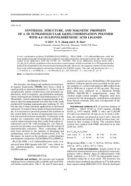
O(4)
C(11) O(5)
N(2) C(16)
fe
N(1) ^
O(11) °(2^) O(9)
O(8) C(20)
C(18)
Fig. 1. Coordination environment of the Gd3+ ion in I. Symmetry codes: (A) -x, 1 - y, -z; (B) -x, 2 - y, -z.
KOOP,3HHAUHOHHAH XHMH3 tom 41 № 5 2015
Fig. 2. The secondary building unit in I. Symmetry codes: (A) — x, 1 — y, — z.
Fig. 3. The 1D chain fragment of I.
Fig. 4. The hydrogen-bonded 3D supermolecular framework of I.
KOOP^HH^HOHHAtf XHMH3 tom 41 № 5 2015
SYNTHESIS, STRUCTURE, AND MAGNETIC PROPERTY
305
Fig. 5. Temperature dependence of XmT and Xm under
Для дальнейшего прочтения статьи необходимо приобрести полный текст. Статьи высылаются в формате PDF на указанную при оплате почту. Время доставки составляет менее 10 минут. Стоимость одной статьи — 150 рублей.