КООРДИНАЦИОННАЯ ХИМИЯ, 2015, том 41, № 4, с. 234-239
УДК 541.49
8-HYDROXYQUINOLINE COORDINATED VANADIUM(V) COMPLEXES WITH SCHIFF BASES: SYNTHESIS, CHARACTERIZATION, AND CRYSTAL STRUCTURES
© 2015 S. S. Qian1, Y. Huo2, Y. T. Ye2, Z. You2, *, and H. L. Zhu1, *
1School of Life Sciences, Shandong University of Technology, ZiBo 255049, P.R. China 2Department of Chemistry and Chemical Engineering, Liaoning Normal University, Dalian, 116029 P.R. China *E-mail: hailiang_zhu@163.com;youzhonglu@126.com Received October 2, 2014
Two new 8-hydroxyquinoline coordinated vanadium(V) complexes, [VO(L1)(L')] (I) and [VO(L2)(L')] (II), where L1 is the dianionic form of 2-[(2-hydroxyethylimino)methyl]-6-methylphenol (H2L1), L2 is the dian-ionic form of N'-(4-oxopentan-2-ylidene)pivalohydrazide (H2L2), L' is the monoanionic form of 8-hydroxy-
quinoline (HL'), were prepared and characterized by elemental analysis, infrared, UV-Vis and 1H NMR spectra, and single crystal X-ray diffraction (CIF files CCDC nos. 1012078 (I) and 1012077 (II)). Complex I crystallizes in the monoclinic space group P2j/n with unit cell dimensions a = 16.120(1), b = 6.9952(5), c = 16.128(1) Â, в = 106.948(2)°, V= 1739.6(2) Â3, Z = 4, GOOF = 1.127, R1 = 0.0464 and wR2 = 0.1172. Complex II crystallizes in the orthorhombic space group Pna2i with unit cell dimensions a = 13.512(2), b = 9.606(1), c = 31.081(4) Â, V = 4034.3(8) Â3, Z = 8, GOOF = 1.044, Rx = 0.0604 and wR2 = 0.1038. The V atoms in the complexes are in octahedral coordination. Thermal stability of the complexes was studied.
DOI: 10.7868/S0132344X15040039
INTRODUCTION
Schiff bases and their metal complexes have received extensive attention in coordination and bioi-norganic chemistry [1—6]. Among the compounds, vanadium complexes have been reported to have interesting biological activities such as normalizing the high blood glucose levels and acting as models of ha-loperoxidases [7—11], as well as catalytic oxidation properties [12—15]. Recently, our research group has reported a few vanadium complexes with biological activities [16—20]. In the present paper, two new 8-hy-droxyquinoline (HL') coordinated vanadium(V) complexes, [VO(L1)(L')] (I) and [VO(L2)(L')] (II), where L1 is the dianionic form of 2-[(2-hydroxyethylimi-no)methyl]-6-methylphenol (H2L1), L2 is the dianionic form of N'-(4-oxopentan-2-ylidene)pivalohy-drazide (H2L2), are presented.
EXPERIMENTAL
Materials and measurements. Commercially available 3-methylsalicylaldehyde, 2-(2-aminoethylami-no)ethanol and pivalohydrazide were purchased from
Sigma-Aldrich and used without further purification. Other solvents and reagents were made in China and used as received. C, H and N elemental analyses were performed with a Perkin-Elmer elemental analyser. Infrared spectra were recorded on a Nicolet AVATAR 360 spectrometer as KBr pellets in the (4000—400) cm-1 region. 1H NMR spectra were recorded on a Bruker 300 MHz instrument. Thermal stability analysis was performed on a Perkin-Elmer Pyris Diamond TGA DTA thermal analyses system.
Synthesis of I. 3-Methylsalicylaldehyde (1.0 mmol, 136 mg) and 2-(2-aminoethylamino)ethanol (1.0 mmol, 104 mg) were dissolved in methanol (20 mL) and stirred at room temperature for 30 min. Then, a methanolic solution (20 mL) of 8-hydroxyquinoline (1.0 mmol, 145 mg) and [VO(Acac)2] (1.0 mmol, 265 mg) was added dropwise to the mixture under stirring. The final mixture was stirred for another 30 min at room temperature to give a yellow solution. The solution was allowed to stand in air for a few days. Yellow block-shaped crystals suitable for X-ray single crystal diffraction were formed at the bottom of the vessel. The iso-
lated product was washed three times with cold methanol, and dried in air. The yield was 49%.
IR data (KBr; v, cm-1): 1627 s, 1563 m, 1497 w, 1458 m, 1433 w, 1379 m, 1310 s, 1266 w, 1225 w, 1101 m, 1048 s, 955 s, 877 m, 825 w, 786 w, 751 s, 641 m, 545 m. 1H NMR (300 MHz, DMSO-d6; 8, ppm): 9.12 (s., 1H), 8.31 (m., 2H), 8.03 (d., 1H), 7.61-7.20 (m., 5H), 7.05 (d., 1H), 6.75 (t., 1H), 5.22 (s., 1H), 4.78 (m., 2H), 4.50 (m., 2H), 1.68 (s., 3H). UV-Vis (acetonitrile; ^max (lgs)): 243 (4.59), 286 (4.10), 335 (3.79), 510 (3.65) nm.
For C19H17N2O4V
anal. calcd., %: C, 58.8; H, 4.4; N, 7.2. Found, %: C, 58.7; H, 4.6; N, 7.1.
Synthesis of II. Pivalohydrazide (1.0 mmol, 116 mg), 8-hydroxyquinoline (1.0 mmol, 145 mg) and [VO(Acac)2] (1.0 mmol, 265 mg) were dissolved in methanol (30 mL) and stirred at room temperature for 30 min to give a brown solution. The solution was allowed to stand in air for a few days. Brown block-shaped crystals suitable for X-ray single crystal diffraction were formed at the bottom of the vessel. The isolated product was washed three times with cold methanol, and dried in air. The yield was 28%.
IR data (KBr; v, cm-1): 1583 s, 1497 s, 1464 s, 1373 s, 1318 s, 1266 m, 1213 w, 1141 w, 1103 s, 1043 s, 960 s, 822 m, 792 w, 748 s, 635 m, 602 m, 533 m, 491 w. 1H NMR (300 MHz, DMSO-d6; 8, ppm): 8.81 (s., 1H), 8.50 (m., 1H), 7.67-7.12 (m., 4H), 2.40 (s., 1H), 2.05 (s., 1H), 1.08 (m., 5H), 0.74 (s., 6H), 0.68 (s., 3H). UV-Vis (acetonitrile; ^max (lgs)): 250 (4.60); 356 (3.98); 517 (3.71) nm.
For C19H22N3O4V
anal. calcd., %: C, 56.0; H, 5.4; N, 10.3. Found, %: C, 56.2; H, 5.6; N, 10.2.
X-ray structure determination. Diffraction intensities for the complexes were collected at 298(2) K using a Bruker D8 VENTURE PHOTON diffractometer with Cu^a (for I) and Mo^a (for II) radiations. The collected data were reduced using the SAINT program [21], and multi-scan absorption corrections were performed using the SADABS program [22]. The structures were solved by direct methods and refined against F2 by full-matrix least-squares methods using the SHELXTL [23]. All of the non-hydrogen atoms were refined anisotropically. H atoms were placed in idealized positions and constrained to ride on their parent atoms. Crystallographic data are listed in Table 1. Selected bond lengths and angles are given in Table 2.
Supplementary material has been deposited with the Cambridge Crystallographic Data Centre (CCDC nos. 1012078 (I) and 1012077 (II); deposit@ccdc. cam.ac.uk or http://www.ccdc.cam.ac.uk).
RESULTS AND DISCUSSION
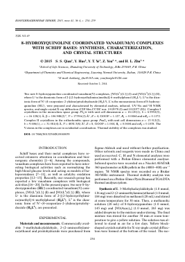
The molecular structure and atom numbering scheme of complex I is shown in Fig. 1a. The V atom is in octahedral coordination, with the phenolate oxygen, imino nitrogen and hydroxyl oxygen of the Schiffbase ligand, and the phenolate oxygen of 8-hy-droxyquinoline ligand in the equatorial plane, and with one oxo oxygen and the pyridine nitrogen of 8-hydroxyquinoline ligand in the two axial positions. The V atom derivates from the least-squares plane defined by the four equatorial atoms by 0.316(1) Â. The distance of V(1)—O(4) is 1.595(2) Â, indicating it is a typical V=O double bond. The bond lengths in the complex are comparable to those observed in the mononuclear oxovanadium(V) complexes with Schiff bases [24, 25]. The angular distortion in the octahedral environment around V comes from the five- and six-membered chelate rings taken by the Schiff base ligand and 8-hydroxyquinoline ligand. For the same reason, the trans angles significantly deviate from the ideal values of 180°. Distortion of the octahedral coordination can be observed from the coordinate bond angles, ranging from 75.4(1)° to 102.9(1)° for the perpendicular angles, and from 157.1(1)° to 173.8(1)° for the diagonal angles.
The molecular structure and atom numbering scheme of complex II is shown in Fig. 1b. The asymmetric unit of the compound contains two independent mononuclear vanadium complex molecules. The V atoms are in octahedral coordination, with the phe-nolate oxygen, imino nitrogen and enolate oxygen of the Schiff base ligand, and the phenolate oxygen of 8-hydroxyquinoline ligand in the equatorial plane, and with one oxo oxygen and the pyridine nitrogen of 8-hydroxyquinoline ligand in the two axial positions. The V atoms derivate from the least-squares planes defined by the equatorial atoms by 0.333(1) Â for V(1) molecule and 0.332(1) Â for V(2) molecule. The distances ofV(1)—O(4) and V(2)—O(8) are 1.59(2) Â, indicating they are typical V=O double bonds. The bond lengths in the complex are comparable to those observed in the mononuclear oxovanadium(V) complexes with Schiff bases [24, 25]. The angular distortion in the octahedral environment around V comes from the five- and six-membered chelate rings taken by the Schiff base ligand and 8-hydroxyquinoline ligand. For the same reason, the trans angles significantly deviate from the ideal values of180°. Distortion of the octahedral coordination can be observed from the coordinate
236 QIAN et al.
Table 1. Crystallographic data and refinement parameters for complexes I and II
Parameter Value
I II
M 388.29 407.34
Crystal shape; color Block; yellow Block; brown
Crystal size, mm 0.18 x 0.18 x 0.17 0.31 x 0.29 x 0.27
Crystal system Monoclinic Orthorhombic
Space group P21/n Pna2i
a, A 16.120(1) 13.512(2)
b, A 6.9952(5) 9.606(1)
c, A 16.128(1) 31.081(4)
ß, deg 106.948(2)
V, A3 1739.6(2) 4034.3(8)
Z 4 8
Pcalcd, g cm-3 1.483 1.341
^(MoÄ"a), mm-1 5.008 0.520
/(000) 800 1696
Measured reflections 9168 24381
Independent reflections 2039 8768
Observed reflections (I > 2ct(/)) 1896 5357
Min and max transmission 0.4659 and 0.4831 0.8555 and 0.8724
Parameters 237 497
Restraints 0 1
Goodness-of-fit on F2 1.127 1.044
R1, wR2 (I> 2a(F))* 0.0464, 0.1172 0.0604, 0.1038
R1, wR2 (all data)* 0.0486, 0.1192 0.1212, 0.1220
Largest peak and deepest hole, e A-3 0.274, -0.623 0.416, -0.388
* R1 = F0 - Fc/F0, wR2 = E w(fO - Fc2)/E w(F02)2]1/2.
KOOP,3HHAUHOHHAH XHMH3 tom 41 № 4 2015
Table 2. Selected bond distances and angles for complexes I and II
Bond d, Â Bond d, Â
V(1)-O(1) V(1)-O(3) V(1)—N(1) V(1)-O(1) V(1)-O(3) V(1)—N(1) V(2)—O(5) V(2)—O(7) V(2)—N(4) 1.880(2) 1.868(2) 2.107(2) I 1.885(3) 1.868(4) 2.053(4)
Для дальнейшего прочтения статьи необходимо приобрести полный текст. Статьи высылаются в формате PDF на указанную при оплате почту. Время доставки составляет менее 10 минут. Стоимость одной статьи — 150 рублей.