КООРДИНАЦИОННАЯ ХИМИЯ, 2015, том 41, № 9, с. 559-565
УДК 541.49
A NOVEL Mn(II) COMPLEX WITH 3-(2,5-DICARBOXYL)-5-CARBOXYLPYRIDINE: SYNTHESIS, CRYSTAL STRUCTURE, AND INTERACTION WITH DNA
© 2015 E. J. Gao*, S. K. Liang, C. Ma, M. C. Zhu, X. Y. Ma, H. T. Jin, F. C. Zhao, and Y. Meng
Department of Coordination Chemistry, Shenyang University of Chemical Technology, International Key Laboratory of Shenyang Inorganic Molecule-Based Chemical, Shenyang, 110142 P.R. China
*E-mail: enjungao@163.com Received February 2, 2015
A novel Mn coordination polymer, |[Mn2(L)6] • 2H2O}n, has been synthesized by the reaction of C4H6MnO4 • 4H2O (manganese acetate) with 3-(2,5-dicarboxyl)-5-carboxylpyridine (L). Elemental analysis, IR spectra, thermal analyses, and X-ray single crystal diffraction (CIF file CCDC no. 1061752) were carried out to determine the composition and crystal structure of the complex. The polymer was characterized by UV spectrum, fluorescence spectrum showing that the complex has the ability of interaction with DNA, Gel electrophoresis assay demonstrated the ability of the complex to cleave the HL-60 DNA (HL-60 DNA, which was extracted by ourselves). Further more, the apoptotic test indicates that the complex has an apop-totic effect on JEKO.
DOI: 10.7868/S0132344X15090029
INTRODUCTION
Recently, researchers have centered an increasing attention on synthesising metal-complexes, rather than individual theory development due to their potential applications as anticancer medications. During the past decades, synthetic coordination chemistry has developed rapidly [1]. Study in this field has provided lots of examples of rationally designed sundry coordination polymers possessing interesting structural motifs and significant properties in catalysis, gas adsorption, magnetism, DNA recognition, and so on [2—6]. There are a large amount of achievements for researchers in seeking drug treatment for cancer. We can hardly control the extended structures, and even the chemical composition of the final products which is one of the key points [7]. Therefore, it's still a long-term challenge to study design and construction of coordination polymers on the basis of the different metal ions and ligands.
There has been substantial interest in the design and investigation of transition-metal anticancer drugs [8—11]. Transition different metal complex of drugs plays an important role in the treatment of the disease. As is well-known, manganese metal is one of the indispensable part of the metal element, carboxylic acids are widely used as ligands as they can easily coordinate with metal by deprotonation. As an important transition-metal element, manganese can also form complexes and some manganese complexes exhibit excellent biological activities [12, 13].
In the present work, based on Mn(II), we synthesized {[Mn2(L)6] • 2H2O}„ by 3-(2,5-dicarboxyl)-5-carboxylpyridine (L). The DNA-cleaving has been investigated by gel electrophoresis. The ability of complex to induce apoptosis is evaluated in JEKO cell line using Annexin V conjugated with FITC and propidium iodide (PI) counterstaining by flow cytometry.
EXPERIMENTAL
Materials and physical measurements. All chemicals purchased were used directly without further purification. IR spectra were recorded in KBr pellets on a Nicolet FT-IR 470 spectrometer in the range of 4000—400 cm-1. UV spectra was recorded on a Shi-madzu UV-240 instrument.
Fluorescence spectra were carried out on a PerkinElmer LS55 fluorescence spectrometer. For all fluorescence measurements, the entrance and exit slits were maintained at 10 and 10 nm, respectively. The samples were excited at 526 nm and its emission observed at 611 nm. The buffer used in the binding studies was 50.0 mM Tris-HCl, pH 6.8-7.3, containing 10.0 mM NaCl. The samples were incubated 4 h at room temperature (20°C) before spectral measurements. The measurement of EB binding to DNA-Mn complex was studied by increasing the concentrations of the complex and measuring the change in fluorescence intensity.
For the gel electrophoresis experiments, HL-60 DNA was treated with the complex in Tris-buffer
Table 1. Crystal data and details of the structure refinement for complex
Parameter Value
Formula weight 716.32
Crystal system Orthorhombic
Space group Pnna
Unit cell dimensions:
a, A 14.5326(18)
b, A 25.046(4)
c, A 7.4669(8)
Z 4
Crystal size, mm 0.2 x 0.2 x 0.2
9 Range for data collection, deg 3.07-27.48
Limiting indices -18 < h < 18, -32 < k < 32,
-9 < l < 9
Reflections collected/unique (R^j) 23699/3109 (0.0677)
Refinement method Full-matrix least-squares
on F2
Data/restraints/parameters 3109/3/216
Goodness-of-fit on F2 1.124
Final R indices (I > 2a(I)) Rx = 0.0408, wR2 = 0.1191
R indices (all data) R1 = 0.0467, wR2 = 0.1265
Largest diff. peak and hole, e A-3 0.476 and -0.601
(50.0 mM Tris-acetate, 18.0 mM NaCl buffer, pH 6.8-7.3), and the contents were incubated for 1.5 h at room temperature. The samples were electrophoresed for 3 h at 90 V on 0.8% agarose gel in Tris-acetate buffer. After electrophoresis, the gel was stained with 1.0 mg/mL EB and photographed under UV light.
Absorption measurements were performed on a Shimadzu UV-2550 double beam spectrophotometer in the range of 200-350 cm-1, using 1 cm path length quartz cuvettes.
The ability of complex to induce apoptosis is evaluated in JEKO cell line using Annexin V conjugated with FITC and propidium iodide (PI) counterstaining by flow cytometry. The JEKO cell in a usable condition were seeded in a 6-well culture plate at 1 x 10⁶ cells per well in a 3 mL culture medium. In 6 and 12 h later the medium including the Mn(II) complex was given. After 12 h (18 h) incubation, cells were gathered, wash cells twice with cold phosphate-buffered saline (PBS) and then resuspend cells in 1x bing-ing buffer at a concentration of 1 x 10⁶ cells/mL. Transfer 100 |L of the solution (1 x 10⁵ cells) to a 5 mL culture tube. Add 5 |L of FITC Annexin V and 5 |L PI. Gently vortex the cells and incubate for 15 min at RT (25°C) in the dark. Add 400 |L of 1 x binding buffer to each tube. Analyze by flow cytometry (Accuri C6, USA) within 1 h.
Synthesis of complex. The concentration of all reagents was 15 mmol/L. For the preparation of the title complex, an aqueous solution (10 mL) containing C4H6MnO4 • 4H2O was added to ligand L aqueous (10 mL). Then the pH of the mixture with KOH solution was adjusted to 5.8. The mixture was stirred for 3 h in air. The clear solution was obtained by filtration under atmospheric pressure. About a month later, the transparent crystals were formed by evaporating the solution at room temperature.
IR (RBr; v, cm-1): 3380 s, 1578 s, 1408 s, 1246 m, 1019 m, 821 s, 780 s.
For C28H18Mn2N2O14
anal. calcd., %: C, 46.99; H, 2.55; N, 3.97. Found, %: C, 46.95; H, 2.53; N, 3.91.
X-ray crystal determination. Structure measurements of the topical complex was performed on a XtaLAB mini X-ray single-crystal diffractometer with MoKa radiation (X = 0.71073 A) at 293 K, and the intensity data were obtained in a range of 3.07° < 9 < < 27.48° at 293 K by using scan technique. A suitable single-crystal of dimensions 0.2 x 0.2 x 0.2 mm was mounted in a glass fiber capillary. A direct method using SHELXS-97 resolved the structure [14, 15]. All non-hydrogen atoms were refined anisotropically. Hydrogen atoms were included in ideal geometrical positions. Crystal data and structure refinement parameters are listed in Table 1. Selected bond lengths and angles are given in Table 2.
Supplementary material for complex has been deposited with the Cambridge Crystallographic Data Centre (CCDC no. 1061752; deposit@ccdc.cam.ac.uk or http:// www.ccdc.cam.ac.uk).
RESULTS AND DISSCUSSION
The crystal unit structure of complex {[Mn2(L)6] • • 2H2O}b was determined by single-crystal X-ray diffraction. The molecular structure of the title complex is shown in Fig. 1. Interestingly, there are two coordinated types of Mn(II) in the complex. The first coordinated mode ofMn(1) is defined by three atoms, namely, four oxygen atoms (o(1), O(1)#1, O(6), O(6)#1) and two nitrogen atoms (N(1), N(1)#1), which reveals O(1) and O(1)#1 coming from 3-(2,5-dicarboxyl) , O(6), N(1) and O(6)#1, N(1)#1 coming from 5-carboxylpyri-dine. The second coordinated type of Mn(2) is ligated to six atoms with four oxygen atoms coming from the ligand (O(2), O(2)#2, O(6), O(6)#2), and the other oxygen atoms coming from the water molecules (O(7) and O(7)#2). The O(1)Mn(1)N(1) angle is 162.63(7)°, the O(6)Mn(1)O(6)#1 angle is 172.27(8)° and the O(1)#1Mn(1)N(1)#1 angle is 162.63(7)°. Also the O(2)#2Mn(2)O(2) angle is 140.60(11)°,
KOOP,3HHAUHOHHAH XHMH3 tom 41 № 9 2015
Table 2. Bond lengths (A) and angles (deg) for complex*
Bond d, A Bond d, A
Mn(1)-O(1) 2.1542(16) Mn(2)-O(2) 2.1389(17)
Mn(1)-O(6) 2.2065(16) Mn(2)-O(6) 2.2334(16)
Mn(1)-N(1) 2.2748(19) Mn(2)-O(7) 2.311(2)
Angle ю, deg Angle ю, deg
O(1)Mn(1)O(1)#x 84.90(10) O(2)#2Mn(2)O(2) 140.60(11)
O(1)Mn(1)O(6) 90.06(6) O(2)#2Mn(2)O(6) 113.62(7)
O(1)#xMn(1)O(6) 95.65(7) O(2)Mn(2)O(6) 92.37(6)
O(6)Mn(1)O(6)#1 172.27(8) O(6)#2Mn(2)O(6) 98.06(8)
O(1)Mn(1)N(1) 162.63(7) O(2)Mn(2)O(7) 84.23(9)
O(1)#xMn(1)N(1) 91.06(7) O(6)Mn(2)O(7) 77.99(8)
O(6)Mn(1)N(1) 73.50(6) O(2)Mn(2)O(7)#2 73.66(8)
O(6)#xMn(1)N(1) 101.26(6) O(6)#2Mn(2)O(7)#2 77.99(8)
N(1)#xMn(1)N(1) 97.50(10) O(6)Mn(2)O(7)#2 162.00(9)
O(7)Mn(2)O(7)#2 110.97(15)
* Symmetry codes: #1 x, -y + 1/2, -z + 1/2; #2 x, -y + 1/2, -z - 1/2.
Fig. 1. The molecular structure of the ligand [Mn2(L)6] • 2H2O (H atoms were omitted for clarity). 4 КООРДИНАЦИОННАЯ ХИМИЯ том 41 № 9 2015
Fig. 2. 1D chain structure of the complex (H atoms were omitted for clarity).
S
a
o U
<4 M
Form II
u^uu
Form III
Form I
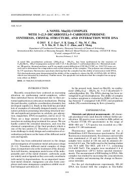
2000 bp
1000 bp 750 bp 500 bp
250 bp
Fig. 3. 2D framework formed by hydrogen-bonding.
Fig. 4. Cleavage of HL-60 DNA in the present of the title complex: marker (lane 0), DNA (lane 1), DNA with diffe
Для дальнейшего прочтения статьи необходимо приобрести полный текст. Статьи высылаются в формате PDF на указанную при оплате почту. Время доставки составляет менее 10 минут. Стоимость одной статьи — 150 рублей.