КОЛЛОИДНЫЙ ЖУРНАЛ, 2013, том 75, № 1, с. 39-44
УДК 541.18
ASSEMBLY AND CHARACTERIZATION OF Ag NANOPARTICLES IN PAM-g-PVA/PVP SEMI-INTERPENETRATING NETWORK HYDROGELS
© 2013 г. Qing-Bo Wei", Yan-Ling Luoé- *, Feng Fu", Lou-Jun Gao", Yrn-Wei Song"
aKey Laboratory of Chemical Reaction Engineering of Shaanxi Province, College of Chemistry &Chemical Engineering,
Yan'an University, Yan'an 716000, P. R. China bKey Laboratory of Macromolecular Science of Shaanxi Province, School of Chemistry and Chemical Engineering,
Shaanxi Normal University, Xi'an 710062, P. R. China Поступила в редакцию 30.09.2011 г.
Polyacrylamide grafted poly(vinyl alcohol)/polyvinylpyrrolidone (PAM-g-PVA/PVP) semi-interpenetrating network (semi-IPN) hydrogels were designed and prepared via a simple free radical polymerization reaction process initiated by a PVA-(NH4)2Ce(NO3)6 redox system. The structure of the PAM-g-PVA/PVP semi-IPNs was characterized by a Fourier transform infrared spectroscopy. The morphologies of PAM-g-PVA/PVP hydrogels and PAM-g-PVA/PVP/Ag nanocomposite hydrogels were examined by scanning electron microscopy and transmission electron microscopy (TEM). The experimental results indicated that the PAM, PVA or PVP chains can efficiently act as stabilizing agents for Ag nanoparticles. TEM investigation of sample morphology showed the presence of nearly spherical-, square- or rectangular-shaped Ag nanoparticles with diameters ranging from 10 to 60 nm. The characteristic surface plasmon resonance band appeared at 390—400 nm as a result of the immobilization of Ag nanoparticles within the hydrogel matrices. The self-assembly of Ag nano-particles and the swelling behavior of the resulting nanocomposites can be controlled and modulated by altering the mole fraction of PVP in the PAM-g-PVA/PVP semi-IPNs.
DOI: 10.7868/S0023291213010151
INTRODUCTION
Over the past decades, metal nanoparticles have attracted considerable attention due to their unique physicochemical properties and potential applications in various fields such as biological catalysts, sensors, optical, magnetic and electronic devices [1—6]. Particularly sliver nanoparticles possess anti-bacterial properties besides the typical characteristics of metal nanoparticles; this combination makes them attractive for some important applications in medicine and immune analysis [7—10]. The performance of nanoparticles is closely associated with the structural characteristics, surface properties, and size distribution, which need to be carefully examined and controlled. Tan et al. prepared well-stabilized and highly dispersed silver nanoparticles by reduction of AgNO3 with NaBH4 in the presence of poly(amidoamine) dendrimer as a template [11]. Jiang et al. reported a rapid and convenient process fabricating silver nanoparticles by microwave high-pressure synthetical technology, using polyacrylamide as a reducing agent and stabilizer [12]. The obtained silver nanoparticles with an average diameter of 66 nm showed narrow distribution and high stability. Microemulsions also represent an important technique to prepare dispersions of nanosized Ag par-
1 Corresponding author. E-mail addresses: luoyanl@snnu.edu.cn or luoyl0401@yahoo.com.cn (YL. Luo).
ticles. Microemulsion structure can precisely control the nanoparticle diameter and stability, and therefore limits the routes of formation of nuclei, growth, gathering and agglomeration of the nanoparticles [13].
Poly(vinyl alcohol) (PVA) is a highly polar, water-soluble and biodegradable polymer [14], and can in situ chemically reduce Ag+. Moreover, the active — OH groups in PVA molecules act as head groups to adsorb Ag+ and guide a directional growth Ag+ ^ Ag [15]. Kacarevic-Popovic et al. reported the reduction of Ag+ ions in a swollen PVA matrix by PVA radicals produced by y-irradiation and fabricated an Ag—PVA hydrogel nanocomposite [16]. Polyvinylpyrrolidone) (PVP) has a good biocompatibility and has been applied for many years as a biomaterial or additive to drug compositions, e.g. as a blood plasma expander and as vitreous humor substitute [17].
The main goal of the study described herein is to design and fabricate stable Ag nanoparticles with narrow size distribution on the basis of the graft PAM-g-PVA/PVP semi-interpenetrating network (semi-IPN) hydrogels as templates. Hydrogels offer a large free space between the swollen cross-linked networks that can act as nanoreactor for the nucleation and growth of the nanoparticles. Functional groups in PAM, PVA or PVP chains can interact with Ag+ ions by a covalent bonding, complex formation or electrostatic interac-
tion. These interactions play an important role in efficient stabilization of the Ag nanoparticles. The PAM-g-PVA/PVP/Ag nanocomposite hydrogels were prepared to study their swelling properties.
2. EXPERIMENTAL SECTION 2.1. Materials and Reagents
The acrylamide (AM), analytical grade (A.G), was supplied by the Shantou Xianhua Chemicals Factory, China. The poly(vinyl alcohol), provided by the Xi'an Chemicals & Glasses Factory, China, served as both a reactant and a reducer, with an average degree of polymerization of 1750. The poly(N-vinylpyrrolidone), pharmaceutical grade, was from the Shanghai WellTone Mater. Technol. Co., Ltd, China, with an average degree of polymerization of 40. Ammonium cerium nitrate ((NH4)2Ce(NO3)6, ACN) as an oxidizer was obtained from the Shanghai Chemical Factory (China), which constituted a redox system together with the PVA. AgNO3, A.G., was supplied by the Shanghai Institute of Fine Chem. Ind. Mater. Sodium hypophosphite (NaH2PO2 • H2O) was supplied by the Beijing Chemical Factory, China. N,N-methylenebi-sacrylamide (NNMBA, A.G.), used as a cross-linker, was purchased from the Tianjin Kermol Chemical Regent Developing Center.
2.2. Preparation of PAM-g-PVA/PVP Grafting Network Hydrogels
PAM-g-PVA/PVP grafting network hydrogels were synthesized by using a free radical polymerization approach. Typically, 0.2 g PVA was dissolved in 10 mL deionized water to form a viscous solution, and then 2g AM monomer and stoichiometric PVP amount were added into the PVA solution. After the stirring proceeded at 50°C for about 10 min, 1 mL of 0.2 mol L-1 ACN solution and 0.02 g (1 wt %, the total weight of the monomer) of NNMBA were added into the reaction mixture under rapid stirring at 50°C for another 5 h. The products were flushed with deionized water for 7 days to remove residues of the unreacted monomers and cross-linking agents. Subsequently, hydrogels were dried in an oven under decreased pressure at 35°C until constant weight was achieved.
2.3. Synthesis of Ag Nanoparticles in PAM-g-PVA/PVP Hydrogel Networks
The synthesis of Ag nanoparticles in the grafting PAM-g-PVA/PVP hydrogel networks was performed according to the following procedure. Dry hydrogels were equilibrated with high-pure water for 3 days. Then the swollen hydrogels were transferred into a beaker containing 50 mL of 0.5 M AgNO3 aqueous solution to diffuse for 1 day. These hydrogels loaded with silver salt were subsequently transferred into a 50 mL
0.5 M NaH2PO2 • H2O aqueous solution to reduce the silver ions into silver nanoparticles at room temperature for 2 h. The grafting PAM-g-PVA/PVP hydrogel networks, containing synthesized silver nanoparticles, are termed below as PAM-g-PVA/PVP/Ag nanocomposite hydrogels. The products were dried in an oven under decreased pressure at 35°C until constant weight was achieved.
2.4. Samples Characterization
The structural characterization of the PAM-g-PVA/PVP and PAA-g-PVA/PVP/Ag systems was carried out on an Equinox 55 Fourier transform infrared spectrometer (Bruker Corp., Germany), using the method of the potassium bromide tableting. The morphological characterization of the samples was performed using a Quanta 200 SEM instrument (Philips-FEI Corp, Netherlands), with an operating voltage of 20 kV. Transmission electron microscopy (TEM) observations were performed on a Hitachi H-600 microscope at an accelerating voltage of 75 kV in order to obtain information on morphologies and sizes ofAg nanoparticles. For UV-vis studies, PAM-g-PVA/PVP/Ag nanocomposite dispersions were filled in a 1 cm path-length quartz cuvette and the spectra were recorded on a TU-1901 UV-vis spectrometer (Purkinje General Instrument, China) using deionized water for background correction.
To understand the swelling of PAM-g-PVA/PVP and PAM-g-PVA/PVP/Ag hydrogels, the dried hydrogels were immersed in an excess amount of deion-ized water at room temperature (25°C) until swelling equilibrium was attained. The equilibrium water content (W») was determined after removing the surface water by blotting with filter paper. The dry weight (W0) was recorded after drying the sample in a vacuum oven at room temperature for 3 days. The equilibrium degree of swelling (Qe) of the samples was calculated from the following equation:
Qe = ( W - W0)/W0. (1)
3. RESULTS AND DISCUSSION 3.1. Fourier Transform Infrared (FTIR) Spectroscopy
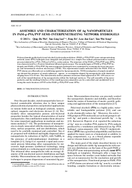
Figure 1 illustrates FTIR spectra of PAM-g-PVA/PVP and PAM-g-PVA/PVP/Ag hydrogels. As shown in Fig. 1a (curve 1), two absorption bands were observed at about 3360 and 3185 cm-1 corresponding to the symmetrical and asymmetrical stretch vibrations of —NH2 groups in AM molecules. The characteristic peaks at 1679 and 1616 cm-1 were assigned to the carbonyl group (-C=O) stretching vibration and the N-H bending vibration of amide groups. In Fig. 1a (curve 2), an absorption peak at 3410 cm-1 was ascribed to -OH stretching vibration present in PVA. A sharp band at 1419 cm-1 was ascribed to a C-O-C
Absorbance (a) Absorbance (b)
4000 3000 2000 1000 4000
Wavenumber, cm-1
3000 2000 1000
Wavenumber, cm-1
Fig. 1. FTIR spectra of (a) AM monomer (curve 1) and PVA polymer (curve 2); and (b): PAM-g-PVA/PVP hydrogel (curve 1) and PAM-g-PVA/PVP/Ag nanocomposite hydrogel (curve 2).
symmetrical stretching mode in acetyl group present in the PVA backbone due to the
Для дальнейшего прочтения статьи необходимо приобрести полный текст. Статьи высылаются в формате PDF на указанную при оплате почту. Время доставки составляет менее 10 минут. Стоимость одной статьи — 150 рублей.