Pis'ma v ZhETF, vol. 93, iss. 7, pp. 446 - 452 © 2011 April 10
Electron-conformational transformations in nanoscopic RyR channels govern both the heart's contraction and beating
A. S. Moskvin+V, A. M. Ryvkin+*, 0. E. Solovyov.a*v, V. S. Markhasin* + Department of Theoretical Physics, Ural State University, 620083 Ekaterinburg, Russia *Institute of Immunology and Physiology, Ural Branch of RAS, 620049 Ekaterinburg, Russia
v Department of Computational Mathematics, Ural State University, 620083 Ekaterinburg, Russia
Submitted 23 December 2010 Resubmitted 21 February 2011
We show that a simple biophysically based electron-conformational model of RyR channel is able to explain and describe on equal footing the oscillatory regime of the heart's cell release unit both in sinoatrial node (pacemaker) cells under normal physiological conditions and in ventricular myocytes under Ca2+ SR overload.
Calcium (Ca2+) dynamics is of a principal importance for functioning of different heart's cells from atrial and ventricular cardiomyocites to sinoatrial node cells (SANC) though the former are responsible for the heart's contraction while the latter for primary heart's pacemaking, respectively [1]. Cardiac contraction in cardiomyocites is activated by an increase in intracellular calcium concentration (Ca2+), most of which comes from a specific calcium cistern of sarcoplasmic reticulum (SR). Ca2+ is released via the ryanodine receptors (RyR) in response to Ca2+ entering the cell via the L-type channels (see Fig. 1). The cardiac type RyR is the common major Ca2+ release channel type in SANC
Cytosol ^^
17 SI
<] SERCA 0
k A
\7
A
Ca;
2+
Ca pumping
up
Jref
Jdiff
2+
Ca diffusion
Cass\
Jre
Ca
nSR
2+
Ca intra-SR diffusion
Ca
jSR
Network SR
I
Junctional SR
»Subspace
M
2+
SR Ca2 clock
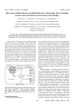
Fig. 1. Schematic illustration of the cell compartments and Ca2+ fluxes in the SR Ca2+ clock toy model (see below Eqs.(7)-(10). SERCA is a sarco/endoplasmic reticulum Ca2+ -ATPase that transfers Ca2+ from the cytosol to the SR
^e-mail: alexandr.moskvin@usu.ru
and ventricular myocytes. It has been experimentally documented in chemically skinned and voltage-clamped SANC, in which effects of voltage-activated sarcolemmal ion currents are excluded, that the isolated SR is capable to spontaneously and rhythmically release Ca2+ via RyRs [2, 3]. These spontaneous, rhythmic, local subsar-colemmal Ca2+ releases (Ca2+ clock), which occur in SANCs, interact somehow with the classic sarcolemmal voltage oscillator (membrane clock [4]). At present there is a general consensus about the importance of Ca2+ oscillator for SANC rate [5], however, an important discussion still remains whether it is a dominant or critical factor for cardiac pacemaker cell functioning. Furthermore, the very existence of the intracellular Ca2+ clock is not captured by the most part of existing essentially membrane-delimited cardiac pacemaker cell numerical models.
Recently, Maltsev and Lakatta[6] have developed a new numerical SANC model (ML-model) featuring interactions of SR-based Ca2+ and membrane clocks to explore novel mechanistic insights into cardiac impulse initiation. They started with a well-known simplified model of the cell structure consisting of four compartments: sub-sarcolemmal space (subspace), cytosol, network SR (nSR), and junctional, or luminal SR (jSR) (Fig. 1). As in most existing models the authors used an effective medium theory, where Ca2+ concentrations in the subspace and in jSR (Cass and Cajsr) are main governing parameters that obey standard reaction-diffusion equations, while RyR gating is usually considered in a simplified manner through a dependence of the release on the Ca2+ concentrations. The ML-model adopted the formulation of cardiac RyR function developed by Shannon et al. [7]) and the Kurata et al. model [8] of primary rabbit SANC. Finally the model
was formulated in terms of a system of 29 first-order differential equations. The isolated SR can indeed operate as a self-sustained Ca2+ oscillator, described by a simple "release-pumping-delay" mechanism: a small spontaneous Ca2+ release from jSR to the subspace occurs as the primary or initiating event. When Cass increases to a sufficient level, it amplifies the Ca2+ release via the mechanism of the Ca2+-induced Ca2+ release (CICR) [1]; this relatively strong, secondary Ca2+ release simultaneously depletes (i.e., resets) jSR. The released Ca2+ is pumped into the nSR. The delay between releases is determined by the Ca2+ pumping rate and Ca2+ diffusion from the subspace to cytosol and also from nSR to jSR. As C&jsr slowly increases, RyRs are restituted, and the next release is ultimately initiated, etc.
The ML-model [6] of coupled oscillators seems to reproduce basically all recently discovered new behavioral details of cardiac pacemaker cell function, however, this phenomenological integrative model ignores many important physiological features of the cardiac cells, in particular, the fine spatiotemporal structure of the Ca2+ release. The model of integrated Ca2+ dynamics does not describe stochastic, locally propagating Ca2+ releases within the subsarcolemmal space. Indeed, RyRs in SANC, as in other cardiomyocites, seem to be arranged in clusters under sarcolemma (Fig. 1) and thus probably form subsarcolemmal calcium release units (CRUs). In this instance, RyRs release Ca2+ into a relatively small volume of subspace where individual jSRs of CRUs approach sarcolemma [1]. Thus a realistic modeling of the Ca2+ oscillator and SANC function should include a stochastic mechanism of a local Ca2+ release generation by CRUs. Furthermore, main assumption of the ML-model [6], that is the Ca2+ released from RyR channels activates the RyR channels as like as the trans-sarcolemmal Ca2+ from the L-type channel in a close apposition, seems to be questionable.
Recently we have applied well-known electron-conformational (EC) model (see, e.g., Ref.[9]) for a single RyR channel [10-12] that was shown to capture important features of the individual and cooperative behaviour of RyRs in ventricular myocytes. The EC model of RyR functioning under Ca2+ stimuli is based on a biophysical adaptation of the well-known theory of photo-induced structural phase transitions, which has been successfully applied to different solids [13]. Hereafter, in the Letter we will show that EC model of RyR channel is able to explain and describe on equal footing a puzzling spontaneous oscillatory regime of the release unit both in SANC under normal physiological
conditions and in ventricular myocytes under Ca2+ SR overload.
The ion-activated RyR channel is a giant (30 x 30 nm) macromolecular protein complex comprising 4 subunits of 565 000 Daltons each [1]. As other ion channels it has a great many of internal electron and conformational degrees of freedom and exhibits remarkable complexities that need to be considered when developing realistic models of ion permeation. Nevertheless, until recently most modelling efforts for RyR channels were focused on a simple "hole in the wall" type model with a set of different (open, closed) states. Our knowledge of molecular mechanisms of RyR channel functioning is limited; hence we are forced to start with the most general "physicists" approach, which is typical for protein biophysics. Such an approach to the modelling of biomolecular system implies its simplifying to bare essentials with guidance from experimental data.
Modelling the RyR we start with a simple and a little bit naive picture of the massive nanoscopic channel like an elastic rubber tube with a varying cross-section governed by a conformational coordinate Q and a light "electronic" plug switched due to Ca2+-RyR binding/unbinding in the subspace [14]. This electronic plug interacts with the conformational coordinate and acts as a trigger to stimulate its change and related channel cross-section/conductivity. In other words, we reduce a large variety of RyR degrees of freedom to only two: a fast and a slow one, conventionally termed as electronic and conformational one, respectively. Both degrees of freedom are implied to be coupled to realize an EC transformation that is the electronic control of the slow conformational motion. Bearing in mind the main function of RyR channels, we assume only two actual electronic RyR states: "open" and "closed", and a single conformational degree of freedom, Q, described by a classical continuous variable. Fig. 2 illustrates the model with a set of representative states of the system.
Change in the electronic and conformational states regulate the main RyR channel function, i.e. determines whether the channel is "open" and permeable for Ca2+ ions or "closed" and impermeable to ions. Hereafter we assume that the conformational variable Q specifies the RyR channel "cross-section" or, more precisely, a permeability for Ca2+, while the dichotomic electronic variable determines its opening and closure. This allows us to describe the Ca2+ flux through the RyR as follows:
JrvR = D(Q)(CajSR - Cass), (1)
if the channel is electronically open, and Jrvr = 0, if it is closed. Here, the permeability coefficient D(Q) reflects the ease with which Ca2+ passes through an open RyR.
OJ
e
i' -
cC
„ cO
w tn\
cC
Q
■Q
Fig. 2. Left panel: Naive "tube-and-plug" model of RyR channel in a closed ground state. Small letters c, o are used for electronically closed and open states, respectively, and capital letters C, O for conformational^ closed and fully open states, respectively. Right panel: Corresponding adiabatic potentials in EC-model of RyR. Vertical arrows point to Ca2+-induced Franck-Condon (FC) electronic transitions, horizontal arrow points to a non-FC tunneling transition, downhill arrows point to a conformational dynamics
Its functional dependence on the conformational coordinate should be one of the essential model assumptions. D(Q) is assumed to be an increasing function
Для дальнейшего прочтения статьи необходимо приобрести полный текст. Статьи высылаются в формате PDF на указанную при оплате почту. Время доставки составляет менее 10 минут. Стоимость одной статьи — 150 рублей.