МОЛЕКУЛЯРНАЯ БИОЛОГИЯ, 2010, том 44, № 4, с. 650-655
МОЛЕКУЛЯРНАЯ БИОЛОГИЯ КЛЕТКИ
UDC 577.24
EXPRESSION PATTERN OF DjPreb GENE DURING THE PLANARIAN Dugesia japonica EMBRYONIC DEVELOPMENT
© 2010 Z. Q. Yuan1, 2, B. S. Zhao2*, J. Y. Zhang2, S. C. Zhang1*
department of Marine Biology, Ocean University of China, 5 Yushan Road, Qingdao 266003, China 2School of Life Sciences, Shandong University of Technology, Zibo 255049, China
Received October 21, 2009 Accepted for publication November 19, 2009
Prolactin regulatory element binding (PREB) protein belongs to the family of WD-repeat proteins which are regulatory and versatile proteins for diverse functions. In this study we have shown the expression pattern of the planarian Dugesia japonica PREB-related gene (DjPreb) during embryonic development by whole-mount in situ hybridization. Genomic analysis reveals that the DjPreb gene consists of two exons and one in-tron. Expression of the DjPreb mRNA was not observed as early as in stage 3 embryos. DjPreb positive signal was first found in stage 4. It is expressed in some embryonic cells in the periphery of the embryo. The number of DjPreb positive embryonic cells grows in stage 5. DjPreb is expressed in the dorsolateral regions and part of the anterior regions in stage 6. In stage 7, DjPreb positive signals are detected in the dorsolateral regions along the A-P axis and from stage 8 to the juvenile stage DjPreb mRNA is strongly expressed not only in the differentiating tissues of the anterior and posterior regions, but also in the parenchyma of the dorsolateral regions, and generates the gradient in the head of the juvenile. These results on the DjPreb expression pattern suggest its potential role in the specification of many cell types; in particular, DjPreb may play an essential role in spatial and temporal regulation during the head and tail formation and the anterior/posterior patterning formation.
Key words: planarian, DjPreb, gene expression, embryogenesis.
WD-repeat proteins are involved in many cellular functions such as signal transduction [1], RNA transcription and processing [2], vesicular trafficking [3], regulation of cytoskeletal assembly [4], cell cycle [5] and cell death [6]. PREB protein contains two potential trans-regulatory PQ-rich domains and three regions of high similarity to the WD-repeat, thus it is considered as a member of the eukaryotic family of WD-repeat proteins [7]. The PREB protein has similarities to a subset of proteins belonging to this family that play the role of gene regulators.
The Preb transcript exhibits a temporal/spatial developmental pattern of expression during murine embryogenesis [8]. Preb is detected in the peripheral nervous system, developing liver, surface ectoderm, Rathke's pouch, axial, and appendicular skeleton. PREB gene is mapped on the human chromosome 2p23, a region of the genome associated with partial trisomy 2p syndrome [9]. PREB transcripts were detected in every human fetal and adult tissue examined; but its expression levels vary significantly among tissues, with very high levels in the pituitary gland, pancreas and the adrenal gland [9—11]. PREB may also play a role during human develop-
* e-mail: zhaobosheng@sdut.edu.cn; sczhang@ouc.edu.cn
ment and the abnormal dosage of this transcription factor may be involved in some of the developmental abnormalities observed in patients with partial tri-somy 2p [9].
Planarians (Platyhelminthes, Tricladida), an invertebrate group well known for their remarkable regenerative ability, retain a population of totipotent stem cells, called neoblasts, throughout their life [12]. Pla-narians have become an excellent model system for studying embryonic development and regeneration biology [13—16]. However, the embryonic development of planarians is still poorly studied. In this study, we have shown the expression pattern of the planarian Dugesia japonica PREB-related gene (DjPreb) during the embryonic development by whole-mount in situ hybridization.
EXPERIMENTAL
Animals and egg capsules. The planarians and egg capsules used belong to a race of D. japonica collected from a fountain in Quanhetou, Boshan, China. The animals were kept in autoclaved tap water at 20°C. Egg capsules were placed in a Petri dish containing autoclaved tap water until reaching the desired stage of de-
5'-UTR
3'-UTR
P1
P3
P2
P4
5'-UTR
3'-UTR
exon [_
] intron
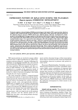
Fig. 1. Primer locations and cloning strategy for the amplification of the planarian (Dugesia japonica) DjPreb gene. Full-length cDNA (a) and intron (b).
a
b
GGGGflCflGGftftGftftGflftTGflTflflTCflTflflTGflftftftGGftflGCGflflTftftflCGftftftTGftflftGCGftftTTCflflTftTGftftftTTGCftTCGTftftft
M K L H R K
TTG^GTCCñTGTTTCCCGTCAGCAññTCññGTGATAññTGCGññTGTTGñAGTGAñTATTGTCAGATñTGGñGñCCññGGñGGCññT LSPCFPSANQU I NflNUEUNIURYGDQGGN TTGGTGATftGCTGGAGGTAATfiATGGfiATTATTCGAGTTTATCfiGAGGTACfiATGATGATCGACCGATGGATTTGGTGgtgagtttg LUIAGGNNGI IRUVQRVNDDRPMDLU ttgaattattgatttatctagtgatgtcacacgccagcagttgtaatgatcattctgtttgtctagTTGCAGCGAGAATTCAACAGC
L Q R E F N S
GGAACACAAGGAGCAGCAGTGATGGATCTCGATGTGTCTCCGTGTCTCGATGAGATGGTGACGGTTTGTGACAATCCAGACGGGAAA
GTQGAAUHDLDUSPCLDEHUTUCDNPDGK
TACGCGCGTATTTGGAGCATTTCTCGAGGCGTGAAAATCACCGAGCTGGCACTGACGAGTATAAGATGCGAGGGAGCCTACAGATTC
VARIWSISRGUKITELALTSIRCEGAVRF
AAACAGTGCAGATTTGCTGCTGCTGGCGGCTGGCTGGCGGGAGGAGGAGGAGGCAGACGAGCTCCATTCTTTCTGTACGCCACTCAC
KQCRFAAAGGWLAGGGGGRRAPFFLYATH
CAGCCAGTTGTGGTGAATGACAAGAACAAATTTGCATTCATGTCTGTCTGGGGCCCTCTCGATCCAGTTGTTGGCACTGATTATCAG
QPUUUNDKNKFAFMSUWGPLDPUUGTDVQ
CTGATTGATGTTGTCGATCTCGGCACGAATTTTGCTGCCTTTGGAACAAGACCAACGAAAATGAGACCAACAGCGCTCTGTACATCC
LIDUUDLGTNFAAFGTRPTKMRPTALCTS
TCATGTGGATTTTTTGTAGGTGTCGGCAGTGGAGAGGGGGCAGTCACTGTTTACAGACTCAACGACCAGACACACAAATTTAGTCGC
SCGFFUGUGSGEGAUTUYRLNDQTHKFSR
ATATACAATCTGCCGAAAGCACACTCATTTTTTGTGACAGACATTGCATTCCTTCCGAGGAATCGTGCATTGAAAACTGGCCATCAA
IVNLPKAHSFFUTDIAFLPRNRALKTGHQ
TTTGAGCTACTGTCAATTGGTGTTGACAACATGTTGCGCTATCATCGCGCCCCTCACAGACCGATTTATTTACGATTGGAAAGATTG
FELLSIGUDNMLRYHRAPHRPIYLR L_E R L
TATAAATTGTTGTCTGTftTTCCTTCTGATATTCATCTGTGATftGAGTTTTCCTCTTGCTGAGCTTGTTTTAftTG|SÂTÂÂ^TATCATC
VKLLSUFLLIFICDRUFLLLSLF*
ATGGTGTTGTTGCGCAAAAAAAAAAAAAAAAAAAAAAAAAAAAAA
Fig. 2. Structure and nucleotide sequence of the planarian (Dugesia japonica) DjPreb, together with the deduced amino acid residue sequence shown underneath. The exons and the 5'- and 3'-UTRs are shown in upper-case letters and the intron - in lowercase letters. The transcription start codon (ATG) is underlined. The asterisk represents the stop codon (TAA). The polyadenyla-tion signal (AATAAA) is boxed.
velopment. Juvenile planarians were collected every day and treated with 2% HCl for 5 min on ice, then fixed on ice for 3 h in the Carnoy's fixative (ethanol : : chloroform : acetic acid, 6 : 3 : 1) and stored in methanol [17].
Fixation. Fresh planarian egg capsules were gently laid on a glass surface, superficially perforated with a 0.5 mm needle, and immediately fixed for 4 h at 4°C in 4% formaldehyde in PBS. After an overnight wash in PBS at 4° C, individual embryos were dissected under the scope with the help of tweezers. After proper dehydration the embryos can be stored in Eppendorf tubes containing 70% ethanol at —20°C till needed.
Genomic DNA extractions. The genomic DNA was extracted as described previously [18]. For total DNA extraction about 100 mg of the planarians were snap frozen in liquid nitrogen, homogenized, and then digested with proteinase K. The protein was removed by phenol and chloroform extraction. The DNA was precipitated by ethanol and dissolved in H2O, then stored in Eppendorf tubes at —20°C until use.
Genomic analysis: identification and sequencing of introns. Genomic clones were created mostly as described previously [18]. Two pairs of primers (namely P1, P2, P3 and P4) were designed according to the sequence of the cloned DjPreb cDNA in order to identify the introns (Fig. 1). Using the extracted genomic
652
YUAN et al.
Fig. 3. Expression patterns of the DjPreb mRNA in Dugesia japonica developing embryos detected by whole-mount in situ hybridization. a — A stage 3 embryo; b — a section of a stage 3 embryo, no signal is observed in the embryo. c — Stage 4 embryo; d — a section of a stage 4 embryo. The DjPreb signal is found in some of the embryonic cells in the periphery of the embryo. e — Stage 5 embryo; /— a section of a stage 5 embryo. DjPreb is expressed in the embryonic pharynx and a cluster of embryonic cell accumulates on the area surrounding it. g — Stage 6 early embryo in dorsal view. Expression of DjPreb is registered in the cells of the anterior and dorsolateral regions. h — A stage 6 later embryo in dorsal view. DjPreb positive signals were detected in the ingrows, the anterior and dorsolateral regions. i — A stage 7 embryo in dorsal view; j — a section of a stage 7 embryo. DjPreb positive signals were detected in the dorsolateral regions. k — A stage 8 embryo in ventral view, DjPreb expression is mainly present in the dorsolateral regions. l — A stage 4 embryo (control) processed and hybridized similarly with the sense probes. No signal is seen in the control. In all images the anterior is to the left. ee — embryonic epidermis; gb — germ band; iym — inner yolk mass; ep — embryonic pharynx. Scale bars represent 100 ^m.
DNA as a template each pair of primers successfully amplified a single fragment. The amplified fragments were then inserted into a cloning vector pMD18-T, which was then transfected into Escherichia coli DH5a, propagated and sequenced. The intron sequences were identified by aligning the amplified frag-
ment nucleotide sequences with the DjPreb cDNA sequences and the GT—AG exon/intron junction nucle-otides.
The PCR primers were designed using the Primer Premier 5.0 software. The PCR primers used were as follows: P1, 5'-GCGAGTAAACGAAATGAAAG-3'; P2, 5'-CACGAGAAATGCTCCAAATA-3'; P3, 5'-ACAGCGGAACACAAGGAGC-3'; P4, 5'-TTCAT-TAAAACAAGCTCAGCAAG-3';
Whole-mount in situ h
Для дальнейшего прочтения статьи необходимо приобрести полный текст. Статьи высылаются в формате PDF на указанную при оплате почту. Время доставки составляет менее 10 минут. Стоимость одной статьи — 150 рублей.
