КОЛЛОИДНЫЙ ЖУРНАЛ, 2007, том 69, № 3, с. 423-425
КРАТКОЕ СООБЩЕНИЕ
УДК 541.182.021:546.59
FORMATION OF SUBMICROMETER-SCALE GOLD NANOPARTICLE AGGREGATES AND THEIR SELF-ORGANIZATION INTO "SUPRACRYSTALS" © 2007 Yonglan Luo
School of Chemistry and Chemical Industry, China West Normal University Nanchong 637002, Sichuan, P. R. China E-mail: luo.yonglan@hotmail.com Поступила в редакцию 01.08.2006 г.
Nearly spherical, submicrometer-scale gold nanoparticle aggregates about 250 nm in diameter are formed directly from a HAuCl^-phenylenediamine aqueous solution at room temperature. As-formed aggregates were characterized by various techniques including scanning electron microscopy, transmission electron microscopy, and X-ray diffraction, and their formation mechanism is also discussed. It is very interesting that "su-pracrystals" can be produced using such gold aggregated nanoparticles.
1. INTRODUCTION
During the past years, nanoparticle research has become the focus of intense research activity and the preparation of nanoparticles have attracted considerable attention from both fundamental and applied points of view due to their unique optical, electrical, and other properties [1].
Gold nanoparticles as the most stable metal nanoparticles will be key materials and building blocks in the 21st century [2]. Since Faraday's first report on the formation of deep red solutions of gold nanoparticles by chemical reduction of an aqueous solution of AuCl- with phosphorus in CS2, the preparation of gold nanoparticles have been widely studied [3, 4]. On the other hand, new properties often emerge from the aggregates that are distinctly different from those of the corresponding isolated nanoparticles and thus the assembly of such nanoparticles into designed architectures is an important goal in materials science [5, 6].
Self-assembly is as an effective and versatile approach to constructing structured system at a molecular level [7], and up to now the construction of nanoparticle aggregates is usually based on such a technique. For example, Jin et al. produced uniform spherical aggregates of magnetic nanoparticles using n-n interactions [8]; Maye et al. constructed spherical assembly of gold nanoparticles using a tetradentate thioether as a mediator [9]; Rotello group used polymers [10] and dendrimers [11] to hold gold nanoparticles together into spherical aggregates via hydrogen-bonding interactions; Goia and Matijevic obtained large gold particles (radius up to 2 ^m) by controlled aggregation of gold nanoparticles with the use of gum Arabic as a steric stabilizer [12]; Velikov et al. fabricated silver spheres (radius up to 1200 nm) by the reduction of AgNO3 with ascorbic acid in an aqueous solution in the presence of a polymeric steric stabilizer [13].
In this letter, a simple method for the preparation of submicrometer-scale gold nanoparticle aggregates about 250 nm in diameter is demonstrated. The formation of such aggregates occurs in a single step upon direct mixing of HAuCl4 and /-phenylenediamine aqueous solutions at room temperature. It is surprising that "supracrystals" can be produced using such aggregates.
2. EXPERIMENTAL PROCEDURES
HAuCl4 was obtained from Aldrich and /-phenylene-diamine from Sanpu Chemical Engineering Corp (Shanghai, China). All reagents were used as received without further purification. All aqueous solutions were prepared with ultrapure water (18 MQ cm-1, Millipore system).
Breefly, submicrometer-scale gold nanoparticle aggregates were prepared as follows: 135 ^l of 0.047 M HAuCl4 aqueous solution were directly mixed with 20 ml of 0.80 mM /-phenylenediamine aqueous solution (i.e., at the /-phenylenediamine-to-gold molar ratio equal to 2.5 : 1) at room temperature. Several minutes later, a large quantity of precipitate was observed. As-prepared precipitate was washed twice with ethanol and water for further characterization.
Samples for scanning electron microscopy (SEM) characterization were prepared by placing large and small amounts of precipitate on an indium-tin oxide (ITO) glass slide and air drying at room temperature, respectively. Sample for transmission electron microscopy (TEM) characterization was prepared by placing a drop of aqueous suspension of such aggregates on carbon-coated copper grid and drying at room temperature. Sample for X-ray diffraction (XRD) analysis was prepared by placing some aggregates on a glass slide and air drying them at room temperature.
424
LUO
\
, , 4000 nm
(a) i-1
> > . / ^ i. , ; i ' L /
if ■:':.■;
(b) 50 nm i-1
(a)
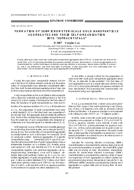
Fig. 2. Typical TEM image of gold nanoparticle aggregates as prepared (a), enlarged TEM image of a single aggregate (b), and SAED pattern of the aggregates (c).
Fig. 1. Low (a) and high (b) magnification SEM images of the film of a large amount of the precipitate on an ITO substrate, indicating the formation of "supracrystals". Inset (b): higher magnification SEM image of the "supracrystals", clearly showing that each aggregate is about 250 nm in diameter and consists of gold nanoparticles.
3. RESULTS AND DISCUSSION
The morphology of films of the precipitates coated on an ITO substrate was characterized by SEM. Figure 1 shows typical SEM images. It is interesting that the sample obtained using a large amount of the precipitate shows the formation of multilayer "supracrystals" as shown in Fig. 1a (low magnification) and Fig. 1b (high magnification). A higher magnification image further reveals that the "supracrystals" consist of nearly spherical nanoparti-cle aggregates about 250 nm in diameter. Also observed are some cracks, which may result from the high surface tension arising during solvent evaporation. The chemical composition of the aggregates was examined by the energy-dispersive X-ray spectroscopy of such a film. The peak of Au was found (other peaks originated from the ITO substrate) (date not shown), indicating the aggregates are gold products.
Typical TEM image of the aggregates thus formed is shown in Fig. 2a, also confirming the formation of nearly spherical particles. Enlarged TEM image of a single particle provides further evidence to support that such particle is an aggregate of small nanoparticles, as shown in Fig. 2b. Figure 2c shows the corresponding selected-area electron diffraction (SAED) pattern obtained by focusing the elec-
tron beam on the aggregates. The SAED pattern reveals that a ring diffraction spot is generated, which demonstrates the crystalline nature of the gold nanoparticle aggregates.
The XRD pattern of aggregates thus formed is shown in Fig. 3. The peaks located at 38.3, 44.5, 64.5, 77.6, and 82.1 degrees are assigned to (111), (200), (220), (311), and (222) faces of an Au crystal, respectively. The observation of the expected diffraction pattern also demonstrates the formation of crystalline gold [14].
HAuCl4 is a powerful oxidant and can be reduced by amine-containing molecules such as o-phenylenediamine [15, 16], polyelectrolyte [17-19] and dendrimer 1[4]. In our study, the spontaneous formation of gold nanoparti-cles can be attributed to the direct redox reaction between HAuCl4 and /-phenylenediamine, because there are no other reducing agents in the system. The formation of nearly spherical gold nanoparticle aggregates can be explained by the mode provided by Matijevic and co-workers [20, 21]. First, HAuCl4 is reduced by /-phenylenedi-amine to form metallic gold atoms. With time, new gold atoms are generated in the system and nucleation occurs as the concentration of gold atoms reaches critical supersaturation, leading to the formation of nuclei. The nuclei grow to nanoscale primary particles by further addition of gold atoms, and then the primary particles form large nearly spherical aggregates. However, the detailed mechanism of the self-organization of such aggregates into "supracrys-tals" is not clear at present time and needs further investigation.
4. CONCLUSION
Direct mixing of HAuCl4 and /-phenylenediamine aqueous solutions at room temperature results in the formation of nearly spherical, submicrometer-scale gold nanoparticle aggregates. Most interestingly, such aggregates can self-organize into multilayer "supracrystals" on a solid substrate. This provides a new methodology for
KOnnOHAHblH XyPHAn TOM 69 < 3 2007
FORMATION OF SUBMICROMETER-SCALE GOLD NANOPARTICLE AGGREGATES
425
Intensity
20, degree
Fig. 3. XRD pattern of the gold nanoparticle aggregates formed on a glass substrate.
preparing submicrometer-scale gold particles appropriate for various applications.
REFERENCES
1. Fendler, J.H., Nanoparticles andNanostructuredFilms, Wiley-VCH, Weinheim, 1998.
2. Daniel, M.C. and Astruc, D., Chem. Rev., 2004, vol. 104, p. 293.
3. Schmid, G., Chem. Rev., 1992, vol. 1709, p. 92.
4. Roucoux, A., Schulz, J., and Patin, H., Chem. Rev., 2002, vol. 102, p. 3757.
5. Lu, Y., Fan, H., Stump, A., Ward, T.L., Rieker, T., and Brinker, C.T., Nature, 1999, vol. 398, p. 223.
6. Breen, T.L., Tien, J., Oliver, S.R.J., Hadzic, T., and Wh-itesides, G.M., Science, 1999, vol. 284, p. 948.
7. Shenhar, R. and Rotello, V.M., Acc. Chem. Res., 2003, vol. 36, p. 549.
8. Jin, J., Lyoda, T., Cao, C.S., Song, Y.L., Jiang, L., Li, T.J., and Zhu, D.B., Angew. Chem. Int. Ed, 2001, vol. 40, p. 2135.
9. Maye, M.M., Chun, S.C., Han, L., Rabinovich, D., and Zhong, C.J., J. Am. Chem. Soc, 2002, vol. 124, p. 4958.
10. Boal, A.K., Ilhan, F., DeRouchey, J.E., Thurn-Albrecht, T., Russell, T.P. and Rotello, V.M., Nature, 2000, vol. 404, p. 746.
11. Frankamp, B., Boal, A.K., and Rotello, V.M., J. Am. Chem. Soc, 2002, vol. 124, p. 15146.
12. Goia, D.V. and Matijevic, E., New J. Chem, 1998, vol. 22, p. 1203.
13. Velikov, K.P., Zegers, G.E., and van Blaaderen, A., Langmuir, 2003, vol. 19, p. 1384.
14. Sun, X., Jiang, X., Dong, S., and Wang, E., Macromol. Rapid Commun., 2003, vol. 24, p. 1024.
15. Sun, X., Dong, S., and Wang, E., Chem. Commun., 2004, p. 1182.
16. Sun, X., Dong, S., and Wang,
Для дальнейшего прочтения статьи необходимо приобрести полный текст. Статьи высылаются в формате PDF на указанную при оплате почту. Время доставки составляет менее 10 минут. Стоимость одной статьи — 150 рублей.