Pis'ma v ZhETF, vol.88, iss. 11, pp.791-794
© 2008 December 10
Observation of the magnetic domain structure in Cuo.47Nio.53 thin films at low temperatures
I. S. Veshchunov, V. A. Oboznov, A. N. Rossolenko, A. S. Prokofiev, L. Ya. VinnikovA. Yu. Rusanov,
D. V. Matveev
Institute of Solid State Physics RAS, 142432 Chernogolovka, Moscow distr., Russia Submitted 28 October 2008
We report on the first experimental visualization of domain structure in films of weakly ferromagnetic Cuo.47Nio.53 alloy with different thickness at liquid helium temperatures. Improved high-resolution Bitter decoration technique was used to map the magnetic contrast on the surface of the films well below the Curie temperature T curie (~60K). In contrast to magnetic force microscopy, this technique allowed visualization of the domain structure without its disturbance while the larger areas of the sample were probed. Maze-like domain patterns, typical for perpendicular magnetic anisotropy, were observed. The average domain width was found to be about 100 nm.
PACS: 75.60.Ch, 75.70.^i
The interplay between superconductivity and ferro-magnetism leads to a number of interesting phenomena [1, 2], which can be utilized in various applications. The interest of investigating the domain structure of weakly ferromagnetic C'ui ¿.Ni,, (x ~ 0.5) alloys, in particular, is caused by their active use in thin film superconductor (S)/ ferromagnet (F) heterostructures. The most promising microelectronic devices based on such heterostructures are basic elements of digital rapid single flux quantum (RSFQ) and quantum logic circuits. Apart from that, using Cui_j,Nij, films as weak ferromagnets in fundamental S/F system properties research looks quite promising, which was confirmed by numerous theoretical and experimental investigations [2-7]. The physical properties of CuNi alloys are relatively well known. The average magnetic moment and the Curie temperature of uniform CuNi alloys decrease linearly with Ni concentration and both approach zero at ~45at.% Ni content. The magnetism of CuNi films is weaker than that in bulk material. Although Cui_j,Nij, films with Ni concentration close to the critical value seem to be good candidates for using them in S/F proximity systems, at the same time they have several disadvantages. One of them is that the film structure is very sensitive to the fabrication conditions. In particular, the homogeneity of sputtered films is not ideal, (at least close to x = 0.5) since there is a tendency of Ni-rich clusters forming [8]. But features of CuNi films domain structure, which have huge influence on the transport and magnetic properties of S/F systems [6] have never been revealed. In this pa-
e-mail: viimik0issp.ac.ru
per we present results of studying magnetic domains in thin films of Cuo,47Nio,53 (hereafter called CuNi) alloy.
In the past decade magnetic force microscopy (MFM) has become a well-established technique for observation of the distribution of magnetic domains with submicron resolution [9-14]. It is widely used, except for the cases when MFM magnetic probe might bring distortions into the scanned image. That can happen, for instance, because of the sample local magnetization change by the probe itself during the scan. It's known that even well below the Curie temperature, the coercive field for thin films of some ferromagnets (CuNi in particular) might become almost zero, so using even magnetically soft MFM probes can disturb the picture of local magnetization when performing scans. Therefore, to study domain structure in such films we used the improved low temperature Bitter decoration technique [15], which also has such additional advantages as high spatial resolution and magnetic sensitivity. The Bitter technique is based on the deposition of fine dispersed magnetic nanoparti-cles, driven by the stray field gradients in the vicinity of magnetic material, at places where the magnetic field is higher. Decoration patterns can be examined by means of scanning electron microscopy (SEM). The patterns provide no information about the magnitude of the magnetization, yet in materials even with low stray fields, Bitter patterns can quickly yield information about the size and shape of domains of various types that might be present.
For our experiments Cuo.47Nio.53 thin films were grown by RF-sputtering in Ar atmosphere of Pai = = 4 • 10-2 mbar on silicon substrates at room temperature. The deposition rate was 0.25 nm/s. The Cu and
IlHCbMa b ?K3T<J> tom 88 Bbin.11-12 2008
791
792
I. S. Veshchunov, V. A. Oboznov, A. N. Rossolenko et a1.
Ni contents in the sputtered films were determined by Rutherford Backscattering (RBS) analysis. It confirmed that the Ni concentration in sputtered films was of the same value as in used CuNi targets.
First, the magnetic properties of Cuo.47Nio.53 films structured in the shape of narrow bridges with thickness dp ranging from 5 to 30 nm were studied by measuring anomalous Hall voltage Fnaii, which is proportional to the film magnetization [16]. Fig. la shows the depen-
10 15 20 25 30 35 40 dF (nm)
10
S*
£ 4
■ dF = 22 nm
У • 1*1.1,1 ^Curie
<Ь) , 1 , 1 , Ч* , *| • .1 , 1 , 1
0 20 40 60 80 100 120 140 T(K)
Figl. (a) Saturation magnetization M as function of the film thickness Af for Cuo.47Nio.53. The dependence has a logarithmic like behavior. The line between experimental points serves to guide an eye. (b) Anomalous Hall voltage VHaii dependence with temperature T for Cuo.47Nio.53 film with dp = 22 nm
dence of Vkaii corresponding to the saturation magnetization of the sample with a particular thickness on the sample thickness. In all cases the applied magnetic field was perpendicular to the film surface. Typical temperature dependence of the Hall voltage for the CuNi film with dF = 22 nm is presented in Fig.lb. For all film thicknesses it appeared to be non linear with a weakly pronounced saturation at low temperatures and tail-like behavior close to the Tcmie- Tfcurie for different sam-
ples was estimated by extrapolating the linear part of the Vkaii (T) dependence as presented in Fig.lb. In order to estimate the field range for the most effective magnetic domain decoration the hysteresis magnetization loops were measured as well. Results of magnetization reversal for Cuo.47Nio.53 film with dj? = 20 nm are presented in Fig.2. The measurements were performed
60 30 0
-30 -60
- Max sweep field (Oe) - 150 Oe „.4* " • 200 Oe / V, - « 250 Oe / . . 300 Oe . " ♦ 700 Oe / J? - v/ ..........
- /
I.I. , 1 , 1
-400
-200 0
Я (Oe)
200
400
Fig.2. Hall voltage VHaii as function of applied magnetic field H for Cuo.47Nio.53 film with dp = 20 nm measured at 4.2 K. Different curves correspond to different maximal sweep fields H as indicated. For all curves H was perpendicular to the film surface. Arrows show the direction of magnetic field sweep
at 4.2 K well below the Tcurie (60 K) of the sample. Several field sweeps were performed with different values of maximum field in the range of 150-700 Oe as shown in Fig.2. The coercive field -ffcoer for those sweeps was found in the range between 50-150 Oe.
Special consideration should be given to the CuNi thin film decoration procedure. The sequence of the entire experiment can be described as follows. The sample was initially cooled in zero magnetic field down to 4.2 K. During the decoration the temperature of the sample increased (of about 3-4 K) up to the decoration temperature Td (the temperature measured by the resistive thermometer in the end of the iron evaporation process). The first series of decoration procedures was performed at magnetic fields H^ec = 100, 250, 300 Oe, on the virgin curve of the hysteresis loop (see Fig.2). For each new field value a new sample (geometrically identical to the previous one) was used. Additional experiment was done to make sure that multiple domain situation occurs after the magnetization switch. For that the magnetic field was gradually increased from 0 to 300 Oe and then swept back to H^ec = ^150 Oe, which corresponds to —-ffcoer for this value of maximum sweeping field, as
Observation of the magnetic domain structure
793
shown in Fig.2. In all cases the applied magnetic field was perpendicular to the sample surface.
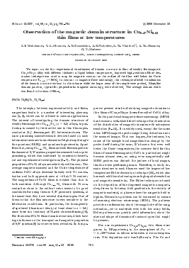
Fig.3 presents the distribution of the iron particles mapping magnetic contrast related to the domain struc-
Место для тонового рисунка
Fig.3. Evolution of the domain structure with external magnetic field applied perpendicular to the film plane: (a) H = 150 Oe, (b) H = 250 Oe, (с) H = 300 Oe, (d) H = ^1500e
ture on the surface of the sample obtained with SEM. Fig.3a, b,c show the domain structure for decoration fields HAeс = 100, 250 and 300 Oe respectively. That, as it has been mentioned before, corresponds to the evolution of the domain state on the virgin curve of the hysteresis loop. At the lowest applied field HAec = 100 Oe the decorated domain structure implies practically demagnetized state on the surface of the sample, which is believed to be perpendicular to the spontaneous magnetization axis. Domains form a maze-like pattern with a typical domain width of about 100 nm. Increasing the decorating field HAec up to 250 Oe as indicated in Fig.3b results in widening of the positive (magnetization is pointed up) domains. Degradation of the pattern quality occurs because of the decrease of the local field gradients on the film surface. The domain structure (i.e. decorated magnetic contrast) almost disappears when approaching HAec = 300 Oe, see Fig.3c. A maze-like domain structure shows up clearly again, as it can be seen from Fig.3d after magnetization switching at the HAe с = Ясоег = ^150 Oe.
The results of investigations reveal several advantages of using CuNi alloys, which justify effectiveness of their utilization in Josephson SFS junctions. Firstly, for this particular ferromagnetic material the exchange
energy is relatively small (Eex/kB ~ 800 K and Tcmi
Для дальнейшего прочтения статьи необходимо приобрести полный текст. Статьи высылаются в формате PDF на указанную при оплате почту. Время доставки составляет менее 10 минут. Стоимость одной статьи — 150 рублей.