ФИЗИОЛОГИЯ РАСТЕНИЙ, 2013, том 60, № 2, с. 240-245
ЭКСПЕРИМЕНТАЛЬНЫЕ СТАТЬИ
УДК 581.1
Physiological and Ultrastructural Responses of Catharanthus roseus Cell Suspension
to Salt Stress1
© 2013 S. Elkahoui*2, Z. Barhoumi**2, N. Djébali***, W. Djebali****, W. Chaïbi****, F. Limam*, A. Smaoui**
* Laboratoire des Substances Bioactives, Centre de Biotechnologie à la Technopole de Borj Cedria, Hammam-Lif, Tunisia ** Laboratoire des Plantes Extrêmophiles, Centre de Biotechnologie à la Technopole de Borj Cedria, Hammam-Lif, Tunisia *** Laboratoire de Physiologie Moléculaire des Plantes, Centre de Biotechnologie à la Technopole de Borj Cedria,
Hammam-Lif, Tunisia
**** Unité de Recherche de Biologie et Physiologie Cellulaires Végétales, Faculté des Sciences de Tunis, Campus Universitaire,
Tunis, Tunisia Received December 22, 2011
Catharanthus roseus (L.) G. Don cell response to salinity was investigated. Seven days after cell treatment with 100 mM NaCl, they showed a decrease in dry weight and an increase in sodium and chloride contents (about 12.4- and 1.5-fold, respectively, in comparison to the control). At the ultrastructural level, NaCl treatment reduced cell size and increased plastid density. In addition, it reduced the starch grain size and their number per plastid; however, starch content was 1.5-fold increased, which was due to the increase in the plastid density. At the ultrastructural level, the applied salinity had no obvious effects, such as swelling or disorganization of plastids except a slight decrease in the stroma electron density. Equally, no deleterious effect was observed on mitochondria except a small increase of their crista volume and matrix electron density. It was shown that, although the relative sensitivity of C. roseus cells to salt stress pointed by the reduction in the dry weight, a decrease in the cell size, and the high accumulation of toxic ions, they preserved the integrity of their plastids and mitochondria.
Keywords: Catharanthus roseus - cell culture - mitochondrion - plastid — salinity
DOI: 10.7868/S0015330313020061
INTRODUCTION
Salinity is an environmental stress that limits plant growth and development. Among various salts existing in saline soils, sodium and chloride are the major ions present at high concentrations, which cause commotion in plants. The response to NaCl excess is complex, and an understanding of the real effects at the cellular level is necessary for elucidation of mechanisms functioning at the level of tissues and whole plants. In fact, some reports support the existence of the correlation between whole plant and cell culture responses to salinity in several species [1]. Some studies indicated that this correlation occurred only if the tolerance of the plant was due predominantly to the cell-based mechanisms [2, 3]. Generally, salinity disturbs plant development through a decrease in the wa-
1 This text was submitted by the authors in English.
2 These authors contributed equally to this work.
Corresponding author. Salem Elkahoui. Laboratoire des Substances Bioactives, Centre de Biotechnologie à la Technopole de Boq' Cedria, B.P. 901, Hammam-Lif 2050, Tunisia. Fax. (+216) 79325728; e-mail. elkahoui@yahoo.fr
ter potential of the root medium; the ions toxicity is due to excessive Na+ and Cl- uptake, and nutrient ion imbalance arises due to the disturbance of essential intracellular ion concentrations [4]. These effects can perturb the physiological and biochemical functioning of the cells and lead eventually to the cell death. The mechanisms of tolerance are those that function to minimize osmotic stress or ion disequilibrium or alleviate the consequent secondary effects caused by salt stress. The response to salinity involves several modifications at the cellular level: morphological, physiological, biochemical, and ultrastructural ones. In fact, there are equivocal reports on the effect of salinity on cells growth [5], and because of the water potential established by the saline solution, the cell turgor declines, and if it decreased below a given limit, the reduction of growth occurred [6]. Under the low water potential, some cells respond by increasing the osmotic potential via the accumulation of compatible os-molytes, such as potassium, sugars, proline, and glycine betaine in the cytosol. These compatible solutes have the capacity to preserve the enzyme activities and have minimal effect on pH or charge balance of the cy-
Table 1. Salt effects on growth, and sodium (Na+) and chloride (Cl-) contents of C. roseus cell suspensions after seven days of 100 mM NaCl treatment
NaCl treatment, mM Growth, g dry wt Na+, mmol/g dry wt Cl , mmol/g dry wt Starch, mg/g dry wt
0 0.67 ± 0.40a 0.17 ± 0.03b 0.37 ± 0.02b 8.00 ± 0.90b
100 0.22 ± 0.03b 2.11 ± 0.04a 0.59 ± 0.01a 13.00 ± 1.10a
Note: Means within the column and marked with different letters are statistically different atp < 0.05 (Duncan multiple rang test).
tosol [7]. At the ultrastructural level, salinity can induce several modifications, such as the development of many vesicles in the cortical cells, as shown in barley and bean [8, 9], vacuolation development, and partial swelling of the endoplasmic reticulum, vesicu-lation and fragmentation of the tonoplast observed in sweet potato [10], rounding of cells and smaller intercellular spaces observed in potato leaves [11]. Equally, salinity induced a substantial modification in mitochondria of the wheat epidermis [12], a decrease in mitochondrial cristae and swelling of these organelles
[10], and a reduction in the number of chloroplasts
[11]. Also, it has been shown that salinity increased the accumulation of lipid bodies and reduced the vacuole size in pea cells [13]. Moreover, these authors also mentioned an increase in the chloroplast size and starch content.
The objective of this work was to evaluate the response of Catharanthus roseus (L.) cell suspensions to NaCl stress at the physiological and ultrastructural levels.
MATERIALS AND METHODS
Cell culture. Catharanthus roseus (L.) G. Don (albino cell lines C20) cell suspension was cultivated in B5 medium [14] supplied with sucrose (58 mM), 2,4-D (4.5 ^M) as the only phytohormone [15], and vitamins. The pH of the medium was adjusted to 5.5 just before autoclaving. Cell suspension was propagated on a rotary shaker (100 rpm) at 24°C in the dark [16]. Cell suspension was subcultured weekly at the dilution 1 : 10 in 250-mL Erlenmeyer flasks containing 50 mL of suspension. For experimental purposes, cells were regularly subcultured during 31 weeks in standard medium. 100 cells were submitted to 0 (control) or 100 mM NaCl during 7 days (on the 32th week).
Measurements of cell growth, ion and starch contents. Cells were collected by filtration and dried at 65°C during four days for dry weight determination. The sodium and chloride analyses were carried out after digestion with 0.5% nitric acid. The sodium concentration was determined by atomic absorption spectrophotometry ("Corning", United States). The chloride content in cells was measured colormetrically ("Haake Buchler", United States). Starch was extracted in a 3% HCl solution and then hydrolyzed to glucose [17]. The starch content was determined using the anthrone method according to Hewitt [18].
Light and electron microscopy. C. roseus cells were fixed at 4°C in glutaraldehyde (2%) in 0.1 M sodium cacodylate buffer (pH 7.4) and postfixed for 1 h in 1% osmium tetroxide in veronal buffer and at the same temperature. The fixed samples were dehydrated in a graded series of ethanol and propylene oxide and embedded in Spurr's epoxy resin ("Oxford Agar", United Kingdom). Semi-thin (1 ^m thickness) sections were prepared using an UltraCut E ultramicrotome ("Re-ichert-Leica", Germany) and were stained with Tolu-idine-O Blue (0.5%, w/v) and Lugol for cell wall and starch staining, respectively. Sections were examined with a photomicroscope ("Leica"). Ultrathin (0.5 ^m thickness) sections were stained with uranyl acetate and lead citrate [19] and observed with a transmission electron microscope (Philips 3015).
Statistical analysis. All experiments were carried out using three replicates for each treatment. The experimental data were subjected to comparison of means using STATISTICA software version 5.1H ("StatSoft", France). Mean values of each treatment were compared using the Duncan multiple range test at 5% probability.
RESULTS
NaCl effect on cell growth, ions accumulation, and starch content
The dry weight of C. roseus cells was decreased by 67% at 100 mM NaCl treatment in comparison to the control (0 mM NaCl) (table 1). The sodium, chloride, and starch concentrations were increased respectively 12.4-, 1.5-, and 1.6-fold in the NaCl-treated cells in comparison to the control (table 1).
NaCl effect on the cell ultrastructure
Light microscopy observations. C. roseus cells treated or not treated with NaCl appeared as agglomerates with a tissues structure and their sizes differed with the age and localization in the tissues (figs. 1a, 1b). In the innermost zone they appeared small, and their arrangement gave the feature of a multiplication zone; however, in the periphery they had large sizes and contained well developed vacuoles (figs. 1a, 1b). NaCl treatment reduced cell sizes in comparison to the control (table 2). In addition, plastid sizes, expressed as the percent of the cell surface, were reduced by NaCl
OH3HOHOrHH PACTEHHH tom 60 № 2 2013
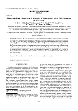
Fig. 1. Microscopic observations of control (a) and 100 mM NaCl-treated (b) C. roseus cells at the seventh day of culture. Note the tissue organization of C. roseus cells in culture and the size and density of plastids (dark spots), which differ between the treated and control cells.
v1 v • ask' i vbi
J * to r' - K " j £ a- - *
<- J-. J*j * -C- ■ V) ^ * ' i,
-ft *
v'CwiImi ■
/ ,5 .J' Cf(¿x i ^r ft . •ft'.'1*-
—a>r r- ,
(a)
(b)
treatment; however, their density was increased 30.
Для дальнейшего прочтения статьи необходимо приобрести полный текст. Статьи высылаются в формате PDF на указанную при оплате почту. Время доставки составляет менее 10 минут. Стоимость одной статьи — 150 рублей.