КООРДИНАЦИОННАЯ ХИМИЯ, 2015, том 41, № 9, с. 533-538
УДК 541.49
SYNTHESES, STRUCTURAL, AND LUMINESCENCE OF TWO COORDINATION POLYMERS
© 2015 L. Lu1, *, W. P. Wu1, A. Q. Ma2, *, B. Xie1, and Y. Wu1
1School of Chemistry and Pharmaceutical Engineering, Sichuan University of Science & Engineering, Zigong, 643000 P.R. China 2Guangdong Medical College, School of Pharmacy, Dongguan, 523808 P.R. China *E-mail: lulusczg@126.com; maqandght@126.com Received February 2, 2015
A organic compound and its lanthanide coordination polymer with chemical formulae HL1, {[La(L1)3(H2O)2] • 2H2O • 2,2'-Bipy} (I), (HL1 = 3-(4-hydroxyphenyl)propanoic acid, 2,2'-Bipy = 2,2'-bipyri-dine) have been synthesized and structurally characterized. Organic compound HL1 shows 1D supramolec-ular chain constructing from intermolecular hydrogen-bonded interaction. Complex II has 1D infinite chain with a [La(L1)3]n dimeric repeat unit with lanthanide in a nine-coordinate environment. Furthermore, the luminescent properties of the two compounds were discussed in detail (CIF files CCDC nos. 1045032 (HL1), 1045033 (I)).
DOI: 10.7868/S0132344X15080034
INTRODUCTION
Coordination polymers have attracted intense attention in recent years because of their intriguing structures and potential applications as functional materials [1—3]. Many efforts has been paid to the rational design of MOFs for the specific needs of applications. A conventional strategy of using long exo-mul-tidentate ligands has been successful to construct frameworks with porosity [4]. Moreover, the flexible carboxylic acids are good candidates for the construction of new coordination polymers as the carboxyl groups can form C—O—M—O cyclic mode with central metal ions, thus, improving the stability of transition metal complexes [4].
Luminescent lanthanide complexes have gained recognition owing to potential applications in biochemistry and materials science [5—7]. Lanthanide ions are excellent luminescent centers and the luminescent properties are influenced by ligands. Compared to first-row transition metals, lanthanides have a larger coordination sphere and more flexible coordination geometry, easily coordinating to various carboxylates and N-containin ligands to form complexes with diverse stereochemistries [8, 9]. Aromatic carboxylic acid, with versatile binding and coordination modes, are widely employed in construction of luminescent lanthanide coordination polymers, which usually exhibit high thermal stability and intense fluorescence because of a large conjugated n-electron system [10—12]. In this report, we employed a flexible carboxylate 3-(4-hydroxyphenyl)propanoic acid (HL1), to build complexes. We anticipated that increase architecture complexity may be introduced by using chelating aromatic N-donor linker and there is an opportunity to
drive the new modes of network assembly required to satisfy the unique constraints imposed by linker geometries. Furthermore, such types of the carboxylate and carboxyl groups of HL1 are always actively involved in H-bonding interactions, which might turn out to be significant structure-controlling factors [12, 13]. The reaction of HL1 with La(III) and the well-known dipyridyl linker of 2,2'-Bipy under mild conditions resulted the formation of a new complex {[La(L1)3(H2O)2] • 2H2O • 2,2'-Bipy} (I), characterized by single crystal X-ray diffraction. Furthermore, the luminescent properties of compounds HL1 and I were discussed in detail.
EXPERIMENTAL
Materials and methods. All reagents were purchased from commercial sources and used as received. IR spectra were recorded with a PerkinElmer Spectrum One spectrometer in the region 4000—400 cm-1 using KBr pellets. TGA was carried out with a Metter-Toledo TA 50 in dry dinitrogen (60 mL min-1) at a heating rate of 5°C min-1. X-ray powder diffraction (XRPD) data were recorded on a Rigaku RU200 dif-fractometer at 60 kV, 300 mA for CuZ"a radiation (X = = 1.5406 A) with a scan speed of 2°C/min and a step size of 0.013° in 29. Luminescence spectra for crystal solid samples were recorded at room temperature on an Edinburgh FLS920 phosphorimeter.
Syntheses of HL1. A mixture of HL1 (0.0149 g), 2,2'-Bipy (0.0220 g), CH3CH2OH (5 mL) and deion-ised water (5 mL) was stirred for 30 min in air. The resulting solution was kept at room temperature for one
Table 1. Crystallographic data and structure refinement information for compound HL1 and I
Parameter Value
HL1 I
Formula weight 166.17 862.64
Crystal system Monoclinic Monoclinic
Space group P2j/c P2l/n
Crystal color Colorless Colorless
a, A 11.275(9) 15.6119(3)
b, A 5.330(4) 9.3468(2)
c, A 14.019(11) 26.9308(5)
P, deg 105.937(14) 106.539(1)
V, A 3 810.1(11) 3767.19(13)
Z 4 4
P calcd g/cm3 1.362 1.521
p., mm-1 0.102 1.201
F(000) 352 1760
9 Range, deg 1.88-25.20 3.21-27.50
Reflection collected 3966 56943
Independent reflections (Rjnt) 3966 (0.0241) 56943 (0.0552)
Reflections with I > 2a(I) 1093 5675
Number of parameters 117 480
R1, wR2 (I> 2ct(I))* 0.0438, 0.1196 0.0371, 0.0767
R1, wR2 (all data)** 0.0594, 0.1388 0.0747, 0.0896
APma« APmin e AT3 0.188, -0.175 0.794, -0.767
* R = S(F0 - Fc)/S(F0), ** wR2 = {S[w(F2 - Fc2)2]/E(FQ2 )2}1/2.
week, the crystals formed were filtered off, washed with water and dried in air.
For C9H10O3 (M = 166.17)
anal. calcd., %: C, 65.05; H, 6.07.
Found, %: C, 65.11; H, 5.92.
Syntheses of I was cattied out by the same synthetic method that for HL1 except that La(NO3)3 ■ 6H2O (0.2 mmol) was introduced into the reactive system.
For C52H52N4O16Cd2 (1213.78)
anal. calcd., %: C, 51.52; H, 5.02; N, 3.25.
Found, %: C, 51.32; H, 5.07; N, 3.04.
IR (KBr; v, cm-1): 3288 v.s, 1904 m, 1534 v.s, 1421 v.s, 1242 v, 1085 m, 826 m,748 m, 523 m.
X-ray crystallography. Single crystal X-ray diffraction analysis of the title compounds were carried out on a Bruker SMART APEX II CCD diffractometer equipped with a graphite monochromated Mo^a radiation (X = 0.71073 Â) by using ® scan technique at
room temperature. Data were processed using the Bruker SAINT package and the structures solution and the refinement procedure was performed using SHELX-97 [14]. The structure was solved by direct methods and refined by full-matrix least-squares fitting on F2. The hydrogen atoms of organic ligands were placed in calculated positions and refined using a riding on attached atoms with isotropic thermal parameters 1.2 times those of their carrier atoms. The hydrogen atoms of lattice water molecule in compounds were located using the different Fourier method. Table 1 shows crystallographic data of HL1 and I. Selected bond distances and bond angles are listed in Table 2. Some H-bonded parameters are listed in Table 3.
Supplementary material for structures HL1 and I has been deposited with the Cambridge Crystallographic Data Centre (nos. 1045032 (HL1), 1045033 (I); depos-it@ccdc.cam.ac.uk or http://www.ccdc.cam.ac.uk).
RESULTS AND DISCUSSION
The adjacent carboxylate groups form a dermic unit through H-bonded interaction (Table 3), then the phenolic groups taking as donor extend the discrete
KOOP^HH^HOHHAtf XHMH3 TOM 41 № 9 2015
Table 2. Selected bond distances (Â) and angles (deg) of structures HL1 and I
Bond d, Â Bond d, Â
C(1)-O(1) C(9)—O(3) La(1)-O(1w) La(1)-O(2w) La(1)-O(5) 1.378(3) 1.290(3) I 2.584(3) 2.576(3) 2.576(3) C(9)-O(2) I La(1)-O(2) La(1)-O(4) La(1)-O(7) 1.203(3) 2.540(3) 2.642(3) 2.539(3)
Angle ro, deg Angle ro, deg
C(1)O(1)H(44) C(9)O(3)H(3^) O(1w)La(1)O(2) O(1w)La(1)O(4) O(2)La(1)O(2w) O(2)La(1)O(5) O(2w)La(1)O(4) O(4)La(1)O(5) O(5)La(1)O(7) 112 (4) 111.00 I 66.95(9) 133.44(9) 138.04(9) 70.56(9) 149.78(9) 49.33(9) 71.46(9) C(1)O(1)H(45) H(4^)O(1)H(45) I O(1w)La(1)O(2w) O(1w)La(1)O(7) O(2)La(1)O(4) O(2)La(1)O(7) O(2w)La(1)O(7) O(4)La(1)O(7) 103(4) 144(6) 72.30(9) 77.43(8) 71.63(9) 86.44(8) 75.23(9) 120.60(8)
Table 3. Hydrogen bonding distances (Â) and angles (deg) for I, II
O(3)-O(2) O(3)-H(3)4-O(2)
I
2.639(7) 174
O(1)'O(1) O(1)-H(4)4-O(1)
II
2.899(2) 178
O(3)-N(2) 2.803(5) O(6)-O(3)w 2.600(1)
O(9)-O(8) 2.7312 O(1)w-O(9) 2.9807
O(1)w-O(3)w 2.7265 O(2)w-O(5) 2.692(2)
O(2)wO(6) 2.828(6) O(3)w-O(4)w 2.682(5)
O(4)wN(1) 2.911(7)
O(3)-H(3)"N(2) 160.00 O(6)-H(6)-O(3)w 138.00
O(9)-H(9)'O(8) 167.00 O(1)w-H(11)-O(9) 157.00
O(1)w-H(11)-O(3)w 159.00 O(2)w-H(21)-O(5) 168.00
O(2)w-H(22)-O(6) 139.00 O(2)w-H(32)-O(4)w 162.00
O(4)w-H(24)-N(1) 155.00
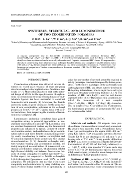
unit into a 1D chain, as shown in Fig. 1. To investigate the complexity on the L1-2,2'-Bipy system, the lanthanum ion was deliberately introduced. A new compound I was obtained. Complex I displays a 1D polymer with [La(L1)3] dimeric repeat units. The asymmetric unit of I consists of one La(III), three L1 anions, two coordinative water molecules, one free 2,2'-Bipy and two lattice water molecules, as shown in Fig. 2a. The
La(III) is nine-coordinate by seven O-donors of six different L1 ligands and two coordinative water molecules. The La(III) can be described as having a cap square antiprism geometry (Fig. 2b) with La-O bond distances ranging from 2.576(3) to 3.006(3) A, all of which are within the range of those observed for other La(II) complexes with oxygen donors. In the polymeric structure of I, the L1 ligand shows n1-^1-^2,
Fig. 1. View of the 1D packing framework directing by weak interactions in HL1.
(a)
(b)
У
O(9) O(3w) \ O(1w) O(8) \ '
O(2w) O(7) O(2)
O(2w) -O(6)
J*.....ë
O(3w)
ff> N
O(3)
O(1w)
Fig. 2. The coordination geometries of the metal centers and the ligands geometries in I (displacement ellipsoids are drawn at the 30% probability level and H atoms are omitted for clarity) (a); SAP geometry surrounding La ion (b); view of the 1D O-C-O-La-O-C-O— chain (c); (d) view of the packing motif directing by weak interactions.
n2-n1-^2 and n0-n0-^1 modes to link two La3+ ions, in which similar with other La-based complexes with carboxylate ligands. The carboxylates of L1 connect La(III) cent
Для дальнейшего прочтения статьи необходимо приобрести полный текст. Статьи высылаются в формате PDF на указанную при оплате почту. Время доставки составляет менее 10 минут. Стоимость одной статьи — 150 рублей.