КООРДИНАЦИОННАЯ ХИМИЯ, 2011, том 37, № 12, с. 938-942
УДК 541.49
SYNTHESIS AND X-RAY CRYSTAL STRUCTURES OF TWO COBALT(III)
COMPLEXES WITH SCHIFF BASES
© 2011 P. Wang, Q. L. Li, Q. Ruan, Y. Q. Su*
Faculty of Chemistry and Chemical Engineering, Yunnan Normal University, Kunming 650092, P.R. China
*E-mail: yongqing_su@163.com Received December 27, 2010
Two new mononuclear cobalt(III) complexes, [Co(MP)2(N3)] (I) and [Co(BP)2]NO3 • 2H2O (II), where MP is 2-methoxy-6-[(2-morpholin-4-ylethylimino)methyl]phenolate and BP is 4-bromo-2-[(2-methylamino-ethylimino)methyl]phenolate, were prepared and structurally characterized by physicochemical methods and single crystal X-ray diffraction. Both complexes crystallize in the monoclinic space group P2i/c. For I: a = 10.3526(18), b = 25.371(4), c = 11.3585(19) А, в = 101.529(8)°, V = 2923.1(8) A3, Z = 4; for II: a = 9.801(2), b = 27.183(3), c = 10.846(2) А, в = 114.269(2)°, V = 2634.2(8) A3, Z = 4. An X-ray structural analysis indicates that in both complexes the Co atoms adopt octahedral coordination. The hindrance effects of the Schiff bases can influence the coordination of the secondary ligands such as azide.
INTRODUCTION
Schiff base complexes of transition metals have attracted much attention in organometallic and coordination chemistry because of their interesting structures and diverse range of applications [1—4]. Cobalt(III) complexes derived from symmetrical and nonsymmetrical Schiff bases have also attracted considerable attention for their im-
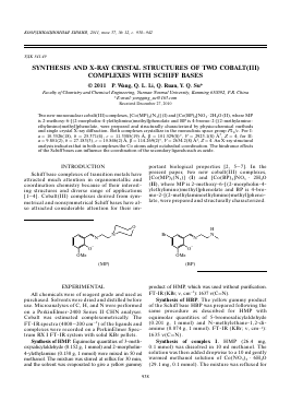
portant biological properties [2, 5—7]. In the present paper, two new cobalt(III) complexes, [Co(MP)2(N3)] (I) and [Co(BP)2]NO3 • 2H2O (II), where MP is 2-methoxy-6-[(2-morpholin-4-ylethylimino)methyl]phenolate and BP is 4-bro-mo-2-[(2-methylaminoethylimino)methyl]pheno-late, were prepared and structurally characterized.
O
OMe
OMe
H
(MP)
(BP)
EXPERIMENTAL
All chemicals were of reagent grade and used as purchased. Solvents were dried and distilled before use. Microanalyses of C, H, and N were performed on a PerkinElmer-2400 Series II CHN analyser. Cobalt was estimated complexometrically. The FT-IR spectra (4000-200 cm-1) of the ligands and complexes were recorded on a PerkinElmer Spectrum RX I FT-IR system with solid KBr pellets.
Synthesis of HMP. Equimolar quantities of 3-meth-oxysalicylaldehyde (0.152 g, 1 mmol) and 2-morpholin-4-ylethylamine (0.130 g, 1 mmol) were mixed in 50 ml methanol. The mixture was stirred at reflux for 30 min, and the solvent was evaporated to give a yellow gummy
product of HMP, which was used without purification. FT-IR (KBr; v, cm-1): 1637 v(C=N).
Synthesis of HBP. The yellow gummy product of the Schiff base HBP was prepared following the same procedure as described for HMP with equimolar quantities of 5-bromosalicylaldehyde (0.201 g, 1 mmol) and N-methylethane-1,2-di-amime (0.074 g, 1 mmol). FT-IR (KBr; v, cm-1): 1635 v(C=N).
Synthesis of complex I. HMP (26.4 mg, 0.1 mmol) was dissolved in 10 ml methanol. The solution was then added dropwise to a 10 ml gently warmed methanol solution of Co(NO3)2 • 6H2O (29.1 mg, 0.1 mmol). The mixture was refluxed for
SYNTHESIS AND X-RAY CRYSTAL STRUCTURES OF TWO COBALT(III) 939
Table 1. Crystal data and structure refinement for complexes I asnd II
Parameter Value
I II
Formula weight 627.58 669.22
Crystal system Monoclinic Monoclinic
Space group P21/c P21/c
Crystal color/shape Brown/block Brown/block
Unit cell dimensions:
a, A 10.3526(18) 9.801(2)
b, A 25.371(4) 27.183(3)
c, A 11.3585(19) 10.846(2)
P, deg 101.529(8) 114.269(2)
Volume, A3 2923.1(8) 2634.2(8)
Z 4 4
P calcd g cm-3 1.426 1.687
Absorption coefficient, mm-1 0.641 3.733
/(000) 1320 1344
9 Range for data collection, deg 2.4-27.7 2.3-24.5
Reflections collected 16914 20774
Independent reflections 6332 5669
Observed reflections (I > 2ct(I)) 4517 2108
Restraints 0 20
Parameters 381 336
Max and min transmission 0.8308 and 0.8459 0.4005 and 0.4322
Goodness-of-fit on F2 1.031 1.024
Final R indices (I> 2ct(I)) 0.0518 0.0779
R indices (all data) 0.0933 0.2331
Largest diff. peak and hole, e A-3 0.779, -0.769 0.726, -0.584
30 min and cooled to room temperature. The resulting deep brown solution was filtered and left undisturbed. After a few days, brown block shaped X-ray diffraction quality single crystals were formed and isolated by filtration. The yield was 17.3 mg (55%).
For C2SH3SN7O6Co
anal. calcd., %: C, 53.6; H, 6.1; N, 15.6; Co, 9.4. Found, %: C, 53.3; H, 6.2; N, 15.7; Co, 9.7.
FT-IR (KBr; v, cm-1): 2028 v(N3), 1621 v(C=N).
Synthesis of complex II was carried out by the same procedure as described for the complex I, only with HMP replaced by HBP (25.7 mg,
0.1 mmol). Complex II is the brown block shaped single crystals. The yield was 21.0 mg (63%).
For C2oH2SN5O7Br2Co
anal. calcd., %: C, 35.9; H, 4.2; N, 10.5; Co, 8.8. Found, %: C, 36.1; H, 4.3; N, 10.3; Co, 9.0.
FT-IR (KBr; v, cm-1): 1381 v(NO3), 1617 v(C=N).
X-ray structure determination. Diffraction quality single crystals of the complexes were mounted on a Bruker AXS SMART diffractometer with a graphite monochromator and MoXa radiation (X = 0.71073 Â). The crystallographic data collection was performed using a multiscan technique at 298(2) K. Data collection and the unit
KOOP^HH^HOHHAtf XHMH3 TOM 37 № 12 2011
940 WANG et al.
Table 2. Selected bond lengths and bond angles for complexes I and II
Bond d, Â Bond d, Â
Co(1)-O(1) Co(1)-N(1) Co(1)—N(4) Co(1)-O(1) Co(1)-N(1) Co(1)—N(3) 1.9170(19) 1.962(2) 2.088(2) I 1.881(7) 1.892(9) 1.896(9) Co(1)—O(3) Co(1)-N(3) Co(1)-N(5) I Co(1)-O(2) Co(1)-N(2) Co(1)-N(4) 1.8835(17) 1.893(2) 1.936(2) 1.880(8) 1.978(10) 1.979(9)
Angle ro, deg Angle ro, deg
O(3)Co(1)N(3) N(3)Co(1)O(1) N(3)Co(1)N(5) O(3)Co(1)N(1) O(1)Co(1)N(1) O(3)Co(1)N(4) O(1)Co(1)N(4) N(1)Co(1)N(4) O(2)Co(1)O(1) O(1)Co(1)N(1) O(1)Co(1)N(3) O(2)Co(1)N(2) N(1)Co(1)N(2) O(2)Co(1)N(4) N(1)Co(1)N(4) N(2)Co(1)N(4) 93.81(9) 83.49(9) 87.96(11) 86.44(8) 92.67(8) 177.54(8) 89.46(8) 94.32(8) I 90.1(3) 93.9(4) 87.6(4) 88.7(4) 85.5(4) 178.5(4) 94.6(4) 92.5(4) O(3)Co(1)O(1) O(3)Co(1)N(5) O(1)Co(1)N(5) N(3)Co(1)N(1) N(5)Co(1)N(1) N(3)Co(1)N(4) N(5)Co(1)N(4) I O(2)Co(1)N(1) O(2)Co(1)N(3) N(1)Co(1)N(3) O(1)Co(1)N(2) N(3)Co(1)N(2) O(1)Co(1)N(4) N(3)Co(1)N(4) 92.84(8) 89.77(9) 171.20(9) 176.15(10) 95.88(10) 85.59(9) 87.82(9) 86.3(4) 94.1(4) 178.5(4) 178.7(4) 93.0(4) 88.7(3) 85.1(4)
cell refinement were performed using the CrysAl-isPRO software [8]. The structures of the complexes were solved by direct method procedures with SHELXS [9] and refined by full-matrix least squares based on F2 with SHELXL [9]. The non-hydrogen atoms were refined with anisotropic factors. The amino and water H atoms in II were located in a difference Fourier map and refined iso-tropically with N—H, O—H and H---H distances restrained to 0.90, 0.85, and 1.37 A, respectively. The remaining hydrogen atoms were positioned geometrically treated as riding on their parent atoms. The data collection and refinement parameters are listed in Table 1. Selected bond lengths and angles are given in Table 2.
Supplementary material for the complxes I and II has been deposited with the Cambridge Crystallographic Data Centre (nos. 803426 (I), 803427 (II); depos-it@ccdc.cam.ac.uk or http://www.ccdc.cam.ac.uk).
RESULTS AND DISCUSSION
The condensation of 3-methoxysalicylaldehyde with 2-morpholin-4-ylethylamine and 5-bromosalicyla-ldehyde with N-methylethane-1,2-diamime in methanol with a 1 : 1 stoichiometric ratio gave two Schiff bases. The Schiff bases react with the Co(NO3)2 • 6H2O and NaN3 in methanol yielding two complexes. It is interesting to note that N3 coordinates with the Co(III) atom in I but not with that in II. A possible explanation is that the hindrance of the morpholine rings in I is larger than that in II, which made one of the morpholine rings in I not coordinating to the Co atom and further led to the coordination of the N3 at the axial position of the octahedral coordination. The Co(II) atom in the starting material is oxidized by air to form Co(III) in two complexes. Elemental analyses are found to be in good
KOOP^HH^HOHHAtf XHMHfl TOM 37 № 12 2011
SYNTHESIS AND X-RAY CRYSTAL STRUCTURES OF TWO COBALT(III)
941
Fig. 1. Molecular structure with atomic numbering for complex I. Hydrogen atoms are omitted for clarity.
agreement with the formular calculated from the X-ray deternination.
The two complexes have v (C=N) absorption at 1621 and 1617 cm-1 which shifts to lower frequencies by 16-18 cm-1 compared to the corresponding Schiff bases, indicating coordintion of the imine N atoms to the Co atoms [10, 11]. A broad band centered at 3617 cm-1 in II is due to the v(OH) in water molecules of crystallization. For both complexes, the phenolic C—O stretching is obtained at about 1277 cm-1. Ligand coordination to the metal center is substantiated by prominent bands appearing at nearly 451 and 372 cm-1, which can be attributed to v(Co-N) and v(Co-O), respectively [12]. The v(N3) band in I appears at 2028 cm-1.
The molecular structure of complex I is shown in Fig. 1. The complex is a neutral mononuclear cobalt compound. The Co atom is hexacoordinat-ed with one phenolate O, one morpholine N, and two imine N atoms from two Schiff base ligands defining the equatorial plane, with one phenolate O atom from a Schiff base ligand, and one azide N atom occupying the axial positions. The coordination geometry is distorted octahedral with the trans and cis angles lying in the ranges 171.2(1)°-177.5(1)° and 83.5(1)°-95.9(1)°, respectively. The Co-N(4)(morpholine) distance is -0.16 Á longer than the average Co-N(imine) distance. The bond lengths and angles are comparable to those observed in other Schiff base cobalt(III) complexes [13-16]. The azide group is almost linear and forms a CoNN angle of 124.9(2)° with the Co atom.
The molecular structure of complex II is shown in Fig. 2. The comp
Для дальнейшего прочтения статьи необходимо приобрести полный текст. Статьи высылаются в формате PDF на указанную при оплате почту. Время доставки составляет менее 10 минут. Стоимость одной статьи — 150 рублей.