КООРДИНАЦИОННАЯ ХИМИЯ, 2010, том 36, № 7, с. 538-543
УДК 541.49
SYNTHESIS, CHARACTERIZATION, AND CRYSTAL STRUCTURES OF TWO SCHIFF BASE ZINC(II) COMPLEXES WITH UREASE INHIBITORY ACTIVITIES
© 2010 D. H. Shi1, 2 * and Z. L. You3
1School of Chemical Engineering, Huaihai Institute of Technology, Lianyungang 222005, P.R. China
2JiangSu-Marine Resources Development Research Institute, Lianyungang 222005, P.R. China
3Department of Chemistry and Chemical Engineering, Liaoning Normal University,
Dalian 116029, P.R. China
* E-mail: dahua_shi@126.com
Received November 19, 2009
Two Schiff base zinc(II) complexes, [ZnBr2L1] ■ 2CH3OH (I) and [ZnBr2L2] (II), where L1 is 4-chloro-2-[(2-piperazin-1-ylethylimino)methyl]phenol and L2 is 4-chloro-2-[(3-diethylamino-propylimino)meth-yl]phenol, were synthesized and structurally characterized. The crystal of I is monoclinic: space group P2i/c, a = 9.831(3), b = 18.680(6), c = 11.879(4) Â, в = 94.660(6)°, V = 2174.3(11) Â3, Z = 4. The crystal of II is monoclinic: space group P21/n, a = 7.2310(14), b = 16.037(3), c = 15.856(3), в = 90.01(3)°, V = 1838.7(6) Â3, Z = 4. The Zn atom in each complex is four-coordinated by one phenolate O and one imine N atoms of the Schiff base ligand and two bromide atoms, forming a tetrahedral coordination. The urease inhibitory activities of the complexes were evaluated.
INTRODUCTION
Urease (urea amidohydrolase, E.C.3.5.1.5) is a nickel-containing metalloenzyme that catalyzes the hydrolysis of urea to form ammonia and carbon dioxide [1, 2]. The enzyme is found in many plants, selected fungi, and a wide variety of prokaryotes [3]. High concentrations of ammonia arising from these reactions, as well as the accompanying pH elevation, have important negative implications in medicine and agriculture [4—6]. The activity of urease through the use of inhibitors can be controlled and thus these negative effects can be counteracted. Recently, we have reported the urease inhibitory activities of some Schiff base cadmium and cobalt complexes [7, 8]. In this paper, two new zinc complexes derived from the Schiff bases, 4-chloro-2-[(2-piperazin-1-ylethyl-imi-no)methyl]phenol (L1) and 4-chloro-2-[(3-diethylami-nopropylimino)methyl]-phenol (L2), were synthesized and evaluated for their urease inhibitory activities.
EXPERIMENTAL
Materials and methods. 5-Chlorosalicylaldehyde, N-(2-aminoethyl)piperazine, and N,N-diethylpropane-1,3-diamine ofAR grade were purchased from Lancaster. Other chemicals and solvents were purchased from the Beijing Chemical Reagent Company and used as received. Elemental analyses for C, H, and N were performed on a PerkinElmer 240C elemental analyzer. The IR spectra
were recorded on a Nicolet AVATAR 360 spectrometer as KBr pellets in the 4000-400 cm-1 region.
Synthesis of L1. To a methanolic solution (20 ml) of 5-chlorosalicylaldehyde (1.0 mmol, 156.6 mg) was added a methanolic solution (20 ml) of N-(2-aminoethyl)pip-erazine (1.0 mmol, 129.0 mg) with stirring. The mixture was stirred for 30 min at room temperature to give a yellow solution. Then the methanol was evaporated to give a yellow precipitate of L1. The yield was 97%.
For C13H18ClN3O
anal. calcd, %: C, 58.3; H, 6.8; N, 15.7. Found, %: C, 58.0; H, 6.8; N, 15.9.
Synthesis of L2. To a methanolic solution (20 ml) of 5-chlorosalicylaldehyde (1.0 mmol, 156.6 mg) was added a methanolic solution (20 ml) of N,N-diethylpropane-1,3-diamine (0.1 mmol, 13.0 mg) with stirring. The mixture was stirred for 30 min at room temperature to give a yellow solution. Then the methanol was evaporated to give a yellow precipitate of L2. The yield was 96%.
For C14H21ClN2O
anal. calcd, %: C, 62.6; H, 7.9; N, 10.4. Found, %: C, 62.8; H, 8.0; N, 10.5.
Synthesis of [ZnBr2L1] • 2CH3OH (I). The Schiff base L1 (0.1 mmol, 26.7 mg) and ZnBr2 (0.1 mmol, 22.5 mg) were mixed and stirred in a methanolic solution (15 ml) for 30 min at room temperature. The mixture was filtered, and the colorless block-shaped crystals ofl suitable for X-ray diffraction, were formed on slow evaporation of the filtrate in air. The yield was 54%.
For C15H26Br2ClN3O3Zn
anal. calcd, %: C, 32.3; H, 4.7; N, 7.5.
Found, %: C, 31.9; H, 5.0; N, 7.3.
Synthesis of [ZnBr2L2] (II). The Schiff base L2 (0.1 mmol, 26.8 mg) and ZnBr2 (0.1 mmol, 22.5 mg) were mixed and stirred in a methanolic solution (15 ml) for 30 min at room temperature. The mixture was filtered, and the colorless block-shaped crystals of II suitable for X-ray diffraction were formed on slow evaporation of the filtrate in air. The yield was 61%.
For C14H21Br2ClN2OZn
anal. calcd, %: C, 34.0; H, 4.3; N, 5.7.
Found, %: C, 34.5; H, 4.3; N, 5.3.
X-ray structure determination. Diffraction intensities for the two complexes were collected at 298(2) K using a Bruker SMART 1000 CCD area-detector with MoZa radiation (X = 0.71073 A). The collected data were reduced with the SAINT program [9], and multiscan absorption corrections were applied using the SADABS program [10]. Both structures were solved by direct methods. The complexes were refined against F2 by full-matrix least-squares methods using the SHELXTL program [11]. All of the non-hydrogen atoms were refined anisotropically. Hydrogen atoms were placed in calculated positions and constrained to ride on their parent atoms. One of the ethyl groups in II is disordered over two distinct sites with occupancies of0.587(2) and 0.413(2). The crystallographic data for both complexes are summarized in Table 1. Selected bond lengths and angles are given in Table 2. Hydrogen bonds are listed in Table 3. Crystallographic data for the complexes have been deposited with the Cambridge Crystallographic Data Centre (nos. 750909 (I) and 750910 (II); deposit@ccdc.cam.ac.uk or http:// www.ccdc.cam.ac.uk).
Urease inhibition assay. The urease inhibitory activities were measured according to the literature method [12]. Generally, the assay mixture containing 25 ^l of jack bean urease (10 kU/l) and 25 ^l of the tested complexes of various concentrations (dissolved in a solution of DMSO : H2O = 1 : 1 (v/v)) was prein-cubated for 1 h at 37°C in a 96-well assay plate. Then 0.2 ml of a 100 mM phosphate buer at pH 6.8 [13] con-
Table 1. Crystallographic data and experimental details for complexes I and II
Vàlue
Parameter
I II
Fw 557.0 494.0
Crystal shape/color Block/colorless Block/colorless
Crystal size, mm 0.32 x 0.28 x 0.27 0.23 x 0.22 x 0.20
Crystal system Monoclinic Monoclinic
Space group P2i/c P21/n
a, A 9.831(3) 7.2310(14)
b, A 18.680(6) 16.037(3)
c, A 11.879(4) 15.856(3)
ß, deg 94.660(6) 90.01(3)
V, A3 2174.3(11) 1838.7(6)
Z 4 4
^(Mo^a), cm-1 4.946 5.827
T J min 0.301 0.347
T J max 0.349 0.389
Pc, g cm-3 1.702 1.784
Measured reflections 16011 11626
Unique reflections and Rint 4650 and 0.0583 4097 and 0.0665
Observed reflections 2942 2453
Restraints 0 21
Parameters 230 213
Goodness of fit on F2 1.005 0.972
Ri (I> 2a(T)) 0.0430 0.0495
wR2 (I > 2a(I)) 0.0841 0.1041
R1 (all data) 0.0845 0.1035
wR2 (all data) 0.0976 0.1240
Large diff. peak and hole, e A-3 0.458, -0.562 0.825, -0.859
KOOP,3HHAUHOHHAH XHMH3 tom 36 № 7 2010
540 SHI,
Table 2. Selected bond lengths and angles for complexes I and II
Bond d, Â Bond d, Â
Zn(1)-N(1) Zn(1)—Br(1) Zn(1)—N(1) Zn(1)—Br(1) ] 2.019(3) 2.3928(9) I 2.019(4) 2.3508(9) [ Zn(1)—O(1) Zn(1)—Br(2) I Zn(1)—O(1) Zn(1)—Br(2) 2.023(3) 2.3787(8) 1.953(4) 2.3598(9)
Angle ю, deg Angle ю, deg
N(1)Zn(1)O(1) O(1)Zn(1)Br(2) O(1)Zn(1)Br(1) O(1)Zn(1)N(1) N(1)Zn(1)Br(1) N(1)Zn(1)Br(2) ] 89.72(13) 97.72(9) 100.86(10) I 93.20(16) 110.30(13) 113.58(13) [ N(1)Zn(1)Br(2) N(1)Zn(1)Br(1) Br(2)Zn(1)Br(1) I O(1)Zn(1)Br(1) O(1)Zn(1)Br(2) Br(1)Zn(1)Br(2) 128.21(11) 119.50(11) 109.24(3) 111.64(11) 112.22(11) 114.12(4)
taining 500 mM urea and 0.002% Phenol Red were added and incubated at 37°C. The reaction time, which was required to produce enough ammonium carbonate to raise the pH of a phosphate buffer from 6.8 to 7.7, was measured by a microplate reader (570 nm) with the end-point, being determined by the color of Phenol Red indicator. The acetohydroxamic acid was used as the standard reference. All the tests were carried out for three times.
YOU
Table 3. Geometric parameters of hydrogen bond for structure I and II*
Contact D-H-A Distance, Â Angle D-H-A,
D-H H-A D-A deg
O(2)H(2)-O(3) I 0.82 1.87 2.692(5) 179
O(3)-H(3)-O(1)#1 0.82 1.80 2.604(4) 168
N(3)—H(3A)-O(2) 0.90 1.91 2.789(5) 164
N(3)-H(3B)-O(3)#2 0.90 2.21 2.958(5) 140
N(3)-H(3B)-Br(2)#3 0.90 2.94 3.519(3) 124
II
N(2)-H(2)-O(1)#4 0.90(5) 1.96(5) 2.857(6) 172(6)
* Symmetry codes: #1 x, y, 1 + z; #2 x, 1/2 --y, 1/2 + z; #4 1/2 + x, 3/2 - y, 1/2 + z. ■ y, -1/2 + z; #3 x, 1/2-
RESULTS AND DISCUSSION
The Schiff bases and two complexes were readily synthesized in methanol solutions. The compounds are stable in air at room temperature, soluble in methanol, eth-anol, and acetonitrile, but insoluble in water.
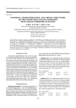
Figures 1 and 2 give perspective views of complexes I and II, respectively. Both complexes are mononuclear zinc compounds. Complex I consists of a zinc complex molecule and two methanol molecules of crystallization.
Fig. 1. Molecular structure of I at 30% probability ellipsoids.
КООРДИНАЦИОННАЯ ХИМИЯ том 36 № 7 2010
C(14)
Br(1)
Fig. 2. Molecular structure of II at 30% probability ellipsoids. Only the major component of the disordered ethyl group is shown.
The Zn atom in each complex has a tetrahedral coordination and is coordinated by one phenolate O atom and one imine N atom of the Schiff base ligand and by two Br atoms. The tetrahedral coordinations are distorted, as evidenced by the bond angles ranging from 89.7(2)° to 128.2(2)° for I and from 93.2(2)° to 114.1(1)° for II. The coordinate bond lengths in both complexes are comparable to each other and also comparable to those observed in other similar Schiff base zinc complexes [14—16].
The molecular packing diagrams for I and II are shown in Figs. 3 and
Для дальнейшего прочтения статьи необходимо приобрести полный текст. Статьи высылаются в формате PDF на указанную при оплате почту. Время доставки составляет менее 10 минут. Стоимость одной статьи — 150 рублей.