КООРДИНАЦИОННАЯ ХИМИЯ, 2012, том 38, № 8, с. 551-556
УДК 541/49
SYNTHESIS, CRYSTAL STRUCTURE, AND DNA INTERACTION AND CLEAVAGE STUDIES OF A NOVEL DINUCLEAR Cu(II) COMPLEX
WITH 3-INDOLYLACETIC ACID © 2012 Y. H. Fan*, L. L. Dong, C. F. Bi, S. B. Liu, X. Zhang, J. Zuo, and Q. Wang
Key Laboratory of Marine Chemistry Theory and Technology, Ministry of Education, College of Chemistry and Chemical Engineering, Ocean University of China, Qingdao, Shandong 266100, P.R. China *E-mail: fyh1959@163.com Received May 16, 2011
A novel dinuclear Cu(II) complex [Cu2(C10H8NO2)4(CH3OH)2] • 2CH3OH (I), where C10H8NO2 is anion of 3-indolylacetic acid, was synthesized and characterized by elemental analysis, IR spectroscopy, 1H NMR and single crystal X-ray diffraction. X-ray crystallography shows that Cu2+ ion is six-coordinated, embedded in a distorted octahedral center. Each Cu2+ ion is coordinated with four carboxylic oxygen atoms from four ligands, one oxygen atom from methanol and the other Cu2+ ion. Each ligand links two Cu2+ ions through carboxylic oxygen atoms, forming a dinuclear Cu(II) complex. The complex forms a two-dimensional layer structure through N—H-Oi intermolecular hydrogen bonds. The interaction of complex I with calf-thymus DNA (CT-DNA) has been explored by electronic absorption spectroscopy, EB (ethidium bromide) displacement experiments, salt effect, and viscosity measurements. All the results indicate that the complex binds to DNA in a partial intercalative mode. Moreover, agarose gel electrophoresis assay demonstrates that the complex possesses the ability to cleave pBR322 plasmid DNA.
INTRODUCTION
A large amount of experiments had elucidated that DNA was the primary intracellular target of anticancer drugs due to the interaction between small molecules and DNA, which caused DNA damage in cancer cells, blocking the division of cancer cells and resulting in cell death [1—3]. Transition metal complexes linked closely to nucleic acids chemistry not only for their diverse applications, such as footprinting agents, sequence specific binding, structural probes and therapeutic agents, but also for their abilities of interacting and cleaving DNA under physiological conditions. Therefore, such complexes are of interest in the development of metal-based anticancer agents [4]. Planar heterocyclic bases complexes have been at the forefront of these investigations because of their unusual electronic properties, diverse chemical reactivity, and peculiar structure, which result in noncovalent interactions with DNA such as intercalation. The intercalation causes changes in the shape of the DNA helix and hinders DNA replication, and RNA transcription [5]. Recent reports have shown that Cu(II) complexes play an important role in the endogenous oxidative DNA damage associated with aging and cancer [6—8]. As a result, investigations on the interaction between complex I with small molecules and DNA are an important preliminary assignment which can provide theoretical basis for drug design, cancer therapy and molecular biology. In this paper, a novel dinuclear Cu(II) complex was synthesized derived from 3-in-
dolylacetic acid, which contains a planar heterocyclic structure. DNA interaction with calf thymus DNA (CT-DNA) was investigated and the achievements of our discussions will be scrutinized in this communication.
EXPERIMENTAL
Reagents and measurement. CT-DNA was biochemical, purchased from Sigma and used as received. A solution of CT-DNA in the Tris-HCl buffer (5 x 10⁻³ mol L-1 Tris-HCl; 5 x 10⁻² mol L-1 NaCl, pH 7.2) gave an absorbance ratio, A260/A280, between 1.8 and 1.9, indicating that the DNA was sufficiently free of protein. This DNA solution was not purified further. The DNA concentration per nucleotide was determined by electronic absorption spectroscopy using the known molar extinction coefficient value of 6600 mol-1 L cm-1 at 260 nm. Other reagents used in this work were of analytical grade.
Elemental analysis (C, H, and N) was performed on a model 2400 PerkinElmer analyzer. Infrared spectrum was recorded as KBr pellets on the Nicolet 170SX spectrometer in the 4000-400 cm-1 region. 1H NMR spectrum was recorded on an AVANCE III (600 MHz) spectrometer. The X-ray diffraction data were collected on an Enraf-Nonius CAD-4 X-ray single-crystal diffractometer. The electronic absorption spectra were recorded on an Avatar 360 FT-IR spectrometer in the range 190-600 nm in methanol solu-
tion. The fluorescence spectra were recorded on an F-4500 fluorimeter.
Synthesis of complex I. The ligand C10H9NO2 (0.175 g, 1 mmol) was dissolved in 5 mL of H2O. Cu(CH3COOH)2 • H2O (0.100 g, 0.5 mmol) dissolved in 10 mL of anhydrous ethanol was added dropwise to the above solution with stirring and refluxed for 3 h to give a green precipitate, which was filtered off, and then dissolved in 15 mL of methanol. The block-shaped crystals were formed in the methanol solution 3 days later.
For C44H4SN4O12Cu2
anal. calcd., %: C, 55.86; H, 4.80; N, 6.06. Found, %: C, 56.09; H, 4.74; N 6.09.
X-ray structure determination. The single crystal with dimensions of 0.48 x 0.42 x 0.40 mm was mounted on an Enraf-Nonius CAD-4 X-ray single-crystal diffractometer. All data were collected at 293(2) K with a graphite monochromatized MoKa radiation (k = 0.71073 A) by using a ®-29 scan mode. The structure was solved by direct methods using SHELXS-97 [9]. The non-hydrogen atoms were defined by the Fourier synthesis method. Positional and thermal parameters were refined by the full matrix least-squares method (on F2) to convergence.
Supplementary material has been deposited with the Cambridge Crystallographic Data Centre (no. 808301; deposit@ccdc.cam.ac.uk or http://www.ccdc.cam. ac.uk).
Electronic spectra study on the interaction between complex I and DNA. The dinuclear complex was dissolved in a mixture solvent of methyl alcohol and Tris-HCl buffer. Absorption titration experiments were carried out by gradually increasing the DNA concentration and maintained the complex concentration constant (5 x 10⁻⁵ mol L-1). Absorbance values were recorded after each successive adding DNA solution and equilibration (~20 min).
EB displacement study on the interaction between complex I and DNA. EB (ethidium bromide) displacement test was carried out by the successive additions of Cu(II) complex to DNA (2 x 10⁻⁶ mol L-1) containing EB (2 x 10⁻⁶ mol L-1) in Tris-HCl buffer. The scanner speed was 1200 nm/min, slit width was 5 nm. These samples were excited at 227 nm. Sterne—Volmer quenching constants were calculated using the given equation: I0/I = 1 + Kq [Q] [10] , where I0 is the emission intensity in the absence of quencher, I is the emission intensity in the present of quencher, Kq is the quenching constant, and [Q] is the quencher concentration.
Salt effect study on the interaction between complex I and DNA. Salt effect was carried out by the successive additions of NaCl to the mixture with concentration of DNA and Cu(II) complex maintained. Fluorescence intensities were recorded in the absence and
presence of NaCl in the mixture solution of the complex and DNA.
Viscosity measurements study on the interaction between complex I and DNA. Viscosity measurements were conducted using an Ubbelodhe viscometer, which immersed in a thermostated water-bath maintained to 298 (±0.1) K. Titrations were performed for the complex which can be introduced into a DNA solution presence in the viscometer. Data were presented as (n/no)1/3 versus the ratio of the concentration of the complex and DNA, where n is the viscosity of DNA in the presence of the complex and n0 is the viscosity of DNA alone.
Cleavage of pBR322 DNA. The cleavage of super-coiled pBR322 DNA (0.1 ^g) by complex I was recorded in Tris-acetic acid-EDTA buffer at pH 7.2 and 310 ± 0.1 K with an incubation time of 4 h, and these samples were analyzed by electrophoresis for 1.5 h at 50 V on a 0.8% agarose gel.
RESULTS AND DISCUSSION
The IR spectrum of complex I displays a strong absorption at 3404 cm-1, which is assigned to v(N—H) of indole ring. The appearance of the bands at 1610.9 and 1420.4 cm-1 are assigned to vas(COO) and vs(COO-), respectively. The absorption at 453.0 cm-1 is attributed to v(Cu-O).
The 1H NMR spectra of complex I are assigned as follows: (DMSO, 600MHz; 8, ppm): 3.647 (2H, s., CH2), 6.984 (1H, d.d., 9-H), 7.077 (1H, d.d., 8-H), 7.232 (1H, d., 3-H), 7.354 (1H, d., 10-H), 7.503 (1H, d., 7-H), 10.915 (1H, s., NH). In the complex I, we can find hydrogen atom of NH still existed, the hydrogen atom of COOH was displaced by the metal ion and a new complex was formed by coordination of copper ion, which is also supported by the IR spectra.
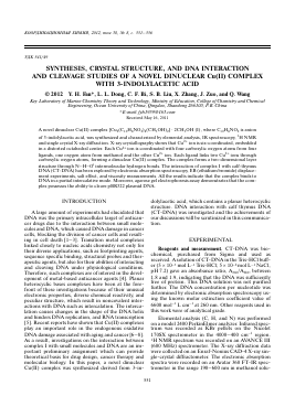
The crystal structure of I is shown in Fig. 1. A summary of the key crystallographic information is given in Table 1, and the selected bond lengths and bond angles are listed in Table 2. The crystallographic structural analysis reveals that complex I is coordinated by four carboxylic oxygen atoms from four ligands, one oxygen atom from methanol and the other Cu2+ ion. The corresponding bond angles of O(3)Cu(1)O(4A) (169.57°) and O(1)Cu(1)O(2A) (169.47) are both less than 180°, and the bond angles O(5)Cu(1)O(1) (89.32°), O(5)Cu(1)O(3) (93.37°), O(5)Cu(1)O(2A) (101.18°), and O(5)Cu(1)O(4A) (96.96°) are less or more than 90°, indicating that the central Cu2+ adopts a distorted octahedron geometry. Four atoms O(1), O(3), O(2A), and O(4A) occupy each vertex of the basal site, while the O(5) and Cu(1A) atoms locate in the apical position of the octahedral structure. As for the bond lengths of Cu(1)-O(1) 1.959, Cu(1)-O(3) 1.961, Cu(1)-O(2A) 1.960, Cu(1)-O(4A) 1.976 A are shorter than that of Cu(1)-O(5) 2.187 A, suggesting that the coordination ability of carboxylic oxygen atoms in the ligand is stronger than that of O(5) in meth-
Fig. 1. Crystal structure of Cu(II) complex. Solvent molecules and all hydrogen atoms were omitted for clarity.
N(2)
Table 1. Crysta
Для дальнейшего прочтения статьи необходимо приобрести полный текст. Статьи высылаются в формате PDF на указанную при оплате почту. Время доставки составляет менее 10 минут. Стоимость одной статьи — 150 рублей.