КООРДИНАЦИОННАЯ ХИМИЯ, 2015, том 41, № 7, с. 409-413
УДК 541.49
SYNTHESIS, CRYSTAL STRUCTURE, AND PRELIMINARY ANTIBACTERIAL ACTIVITY OF OXOVANADIUM(V) COMPLEX WITH HYDRAZONE LIGAND
© 2015 W. Li1, *, X. Han2, and Y. Ding3
department of Hepatobiliary and Pancreatic Surgery, the Second Affiliated Hospital of Dalian Medical University,
Dalian, 116029 P.R. China
2Liaoning Grain and Oil Inspection and Monitoring Department, Shenyang, 110032 P.R. China
3Department of Otorhinolaryngology, the Second Affiliated Hospital of Dalian Medical University, Dalian, 116023 P.R. China
*E-mail: liwei_dlmu@126.com
Received January 12, 2015
A new mononuclear oxovanadium(V) complex, [VOL(OMe)(MeOH)] (I), where L is the dianionic form of (1#-indol-3-yl)acetic acid [1-(2-hydroxynaphthalen-1-yl)methylidene]hydrazine, has been synthesized and characterized by physico-chemical methods and single crystal X-ray determination (CIF file CCDC no. 858861). The complex crystallizes in the triclinic space group PI with unit cell dimensions a = 10.117(1), b = 10.609(2), c = 11.454(2) A, a = 99.228(2)°, P = 112.250(2)°, y = 100.561(2)°, V = 1082.3(3) A3, Z = 2, Rj = 0.0512, and wR2 = 0.1045. The V atom in complex I is in an octahedral coordination. Thermal analysis and preliminary antibacterial activity of complex I were studied.
DOI: 10.7868/S0132344X1507004X
INTRODUCTION
Schiff base complexes play an important role in the development of coordination chemistry related to catalysis and enzymatic reactions, magnetism and molecular architectures [1—4]. Hydrazone compounds, bearing —CH=N—NH—C(O)— functional groups, are a special kind of Schiff bases, which have extensive biological properties [5—8]. In recent years, some metal complexes with hydrazone ligands have been reported [9, 10]. But when compared to the general Schiff base complexes, the number of hydrazone-type complexes is much less. Considering that vanadium ion plays important role in biological processes [11—13], the present work designed, synthesized and studied on the antibacterial activity of a new oxovanadium(V) complex [VOL(OMe)(MeOH)] (I) with the hydrazone ligand (1#-indol-3-yl)acetic acid [1-(2-hydroxy-naphthalen-1-yl)methylidene]hydrazine (H2L).
N ^OH
(ВД
EXPERIMENTAL
Materials and measurements. Commercially available 2-hydroxy-1-naphthyaldehyde, (1H-indol-3-yl)acetic
acid hydrazide and VO(Acac)2 were purchased from Lancaster and used without further purification. Other solvents and reagents were made in China and were used as obtained. C, H and N elemental analyses were performed with a PerkinElmer 240C elemental analyser. IR spectra were recorded on a Nicolet AVATAR 360 spectrometer as KBr pellets in the 4000—400 cm-1 region. Thermal analysis was carried out on PerkinElm-er Pyris Diamond TG-DTA thermal analyses system at a temperature range of 30-1000°C.
Synthesis of H2L. The Schffbase ligand H2L was prepared by the condensation of 2-hydroxy-1-naphthalde-hyde (1.0 mmol, 172 mg) with (1#-indol-3-yl)acetic acid hydrazide (1.0 mmol, 189 mg) in methanol (20 cm3) at room temperature. The yield was 78%.
For C21H!7N3O2
anal. calcd., %: C, 73.4; H, 5.0; N, 12.2. Found, %: C, 73.6; H, 5.0; N, 12.1.
Synthesis of complex I. A methanol solution (5 cm3) of VO(Acac)2 (0.1 mmol, 26.5 mg) was added to a methanol solution (10 cm3) of H2L (0.1 mmol, 34.3 mg) with continuous stirring. The mixture was stirred for 20 min at room temperature to give a deep brown solution. Upon keeping the solution in air for several days, brown block-like single crystals suitable for X-ray diffraction were deposited at the bottom of the vessel. The isolated product was washed three times
Table 1. Crystallographic data and structure refinement for complex I
Parameter Value
Crystal shape/colour Block/brown
Crystal size, mm 0.32 x 0.30 x 0.27
Crystal system Triclinic
Space group P1
a, A 10.117(1)
b, A 10.609(2)
c, A 11.454(2)
a, deg 99.228(2)
P, deg 112.250(2)
Y, deg 100.561(2)
V, A3 1082.3(3)
Z 2
MMoKa), A 0.71073
^(MoKa), cm-1 0.499
^min/^max 0.8567/0.8771
Measured reflections 8147
Independent reflections 4566
Observed reflections (I > 2ct(I)) 3102
Number of refinement parameters 296
Restraints 2
Goodness of fit on F2 1.035
R1, wR2 (I> 2ct(T))* 0.0512, 0.1045
R1, wR2 (all data)* 0.0847, 0.1170
-- SjjiO! - i^cii/Si^oi, wR2 = [Sw(fO2 - Fc2)2/Sw(Fo2)2]1/2, w = [ct2(Fo)2 + (0.0449(F2 + 2Fc2)/3)2 + 0.2273(F2 + 2Fc2)/3]-
with cold methanol and dried in a vacuum over anhydrous CaCl2. The yield was 53%.
For C23H22N3O5V (Fw = 471.4)
anal. calcd., %: C, 58.6; H, 4.7; N, 8.9.
Found, %: C, 58.4; H, 4.6; N, 9.0.
X-ray structure determination. Diffraction intensities for complex I were collected at 298(2) K using a Bruker APEX II area-detector with MoKa radiation. The collected data were reduced using SAINT program [14], and multiscan absorption corrections were performed using SADABS program [15]. The structure was solved by direct methods and refined against F2 by full-matrix least-squares methods using SHELXTL program [16]. All of the non-hydrogen atoms were refined anisotropically. H(5) and H(3) atoms were located from a difference Fourier map and refined isotropically with O-H and N-H distances restrained to 0.85(1) and 0.90(1) A, respectively. The remaining hydrogen atoms in the complex were placed in calculated positions and constrained to ride on their parent atoms. The crystallographic data for
the complex are summarized in Table 1. Selected bond lengths and angles are given in Table 2.
The supplementary crystallographic data for complex I has been deposited with the Cambridge Crystallographic Data Centre (CCDC no. 858861; deposit@ccdc.cam.ac.uk or http://www.ccdc. cam.ac.uk).
Table 2. Selected bond lengths (A) and bond angles (deg) for I
Bond d, A Bond d, A
V(1)-O(1) V(1)-O(3) V(1)-O(5) 1.853(2) 1.574(2) 2.466(2) V(1)-O(2) V(1)-O(4) V(1)-N(1) 1.953(2) 1.771(2) 2.112(2)
Angle ro, deg Angle ro, deg
O(3)V(1)O(4) O(4)V(1)O(1) O(4)V(1)O(2) O(3)V(1)N(1) O(1)V(1)N(1) O(3)V(1)O(5) O(1)V(1)O(5) N(1)V(1)O(5) 102.2(1) 103.5(1) 94.0(1) 94.2(1) 81.7(1) 176.4(1) 82.0(1) 85.4(1) O(3)V(1)O(1) O(3)V(1)O(2) O(1)V(1)O(2) O(4)V(1)N(1) O(2)V(1)N(1) O(4)V(1)O(5) O(2)V(1)O(5) 101.5(1) 100.4(1) 148.2(1) 161.2(1) 74.0(1) 77.7(1) 76.0(1)
KOOP^HH^HOHHAtf XHMH3 tom 41 № 7 2015
RESULTS AND DISCUSSION
Facile condensation of 2-hydroxy-1-naphthyalde-hyde with (1#-indol-3-yl)acetic acid hydrazide in an
The crystals of I are soluble in most polar organic solvents, such as methanol, ethanol, DMF, and DMSO. Elemental analysis is in good agreement with the chemical formula proposed for the complex. In dry methanol, complex I behaves as a non-electrolyte as reflected in its AM value of 31 fi-1 cm2 mol-1 [17].
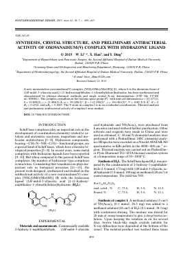
Figure 1 gives perspective view of the complex together with the atomic labeling system. X-ray crystallography reveals that the compound is a mononuclear vanadium complex. The V atom in the complex is in an octahedral coordination, with the three donor atoms of L and one methanolato O atom defining the equatorial plane, and with one oxo O atom and one methanol O atom occupying the axial positions. The distance V(1)-O(3) is 1.574(2) Á, indicating it is a typical V=O bond. The coordinate bond lengths in the complex are comparable to those observed in mono-nuclear oxovanadium complexes with octahedral coordination [18-20]. The distortion of the octahedral
1 : 1 molar ratio furnished the ligand H2L. Complex I was formed in methanol solution containing equimo-lar quantities of H2L and VO(Acac)2:
coordination can be observed from the coordinate bond angles, ranging from 73.96(8)° to 103.50(8)° for the perpendicular angles, and from 148.25(9)° to 176.39(9)° for diagonal angles. The displacement of the V atom from the equatorial plane towards the axial oxo O atom is 0.328(1) Á. The formation of the coordinate bonds with the V atoms, together with the delo-calization, lead to the planarity of the naphthylene ring and the V(1)-N(1)-N(2)-C(12)-O(2) chelate ring. The naphthylene ring forms dihedral angles of 18.2(3)° and 69.8(3)° with the V(1)-N(1)-N(2)-C(12)-O(2) chelate ring and the indole ring, respectively. In the crystal structure, adjacent two molecules are linked through intermolecular O(5)-H(5)—N(2) hydrogen bonds (O(5)-H(5) 0.85(1), H(5)-N(2)' 1.88(1), O(5)---N(2)i 2.727(3) Á, O(5)-H(5)-N(2)' 177(3)°; symmetry code for i: 1 - x, -y, 1 - z), forming a dimer. The dimers are further linked through intermolecular N(3)-H(3)---O(5) hydrogen bonds
Fig. 1. The structure of complex I, showing the atom-numbering scheme. Displacement ellipsoids are drawn at the 30% probability level and H atoms are shown as small spheres of arbitrary radii.
KOOP^HH^HOHHAtf XHMH3 TOM 41 № 7 2015
Fig. 2. Molecular packing of I, viewed down the z axis.
(N(3)-H(3) 0.90(1), H(3)-O(5)u 2.19(1), N(3)-O(5)u 3.084(3) A, N(3)-H(3)-O(5)u 176(3)°; symmetry code for ii: -x, -y, 1 - z), to form 1D chains running along the x axis, as shown in Fig. 2.
The medium and broad absorption centered at 3320 cm-1 is assigned to the vibration of the hydroxyl groups of the coordinate methanol molecule. The weak and sharp absorption at 3135 cm-1 is assigned to the vibration of the N-H group. The typical strong v(C=O) absorption band of the free hydrazone ligand is absent in complex I, indicating the enolisation of the amide functionality group. The strong absorption band at 1605 cm-1 is assigned to the vibration of the
Table 3. Antibacterial screening results
Zone of inhibition, mm
Compound E. coli P. aeruginosa B. subtilis S. aureus
H2L 15 ± 3.3 4 ± 3.3 * *
Complex I 20 ± 5.8 13 ± 3.3 5 ± 3.3 9 ± 3.3
Penicillin G 31 ± 3.3 27 ± 3.3 29 ± 5.8 25 ± 3.3
DMSO - - - -
* Indicates that the bacteria are resistant to the compound.
-I 140
200 400 600 800 1000 Temperature, °C
Fig. 3. TG—DTA curves of complex I.
azomethine group, v(C=N) [21]. The band indicative of the V=O vibration is at 960 cm-1 [22].
Differential thermal (DTA) and
Для дальнейшего прочтения статьи необходимо приобрести полный текст. Статьи высылаются в формате PDF на указанную при оплате почту. Время доставки составляет менее 10 минут. Стоимость одной статьи — 150 рублей.