ОПТИКА И СПЕКТРОСКОПИЯ, 2014, том 117, № 3, с. 401-405
СПЕКТРОСКОПИЯ КОНДЕНСИРОВАННОГО СОСТОЯНИЯ
УДК 543.42
EFFECTS OF 1064 nm LASER ON THE STRUCTURAL AND OPTICAL PROPERTIES OF NANOSTRUCTURED TiO2 THIN FILM
© 2014 г. W. Aslam Farooq, M. Atif, Syed Mansoor Ali, Amanullah Fatehmulla, and M. Aslam
Department of Physics and Astronomy, College of Science P.O. Box 2455, King Saud University Riyadh, Saudi Arabia
E-mail: wafarooq@hotmail.com Received November 11, 2013
TiO2 thin film has been widely used as photoelectrode in dye sensitized solar cells. It can also be used in quantum dots synthesized solar cells. Study of its effects in different spectrum of light is important for its use in solar cells. We have reported effects of 1064 nm laser on the surface morphology, structural and optical properties of nanostructured TiO2 thin film deposited on glass substrates using sol gel spin coating technique. Q-switched Nd : YAG pulsed laser at various power densities is used in this study. Surface morphology of the film is investigated using X-ray diffraction and atomic force microscopy technique. The XRD pattern of as deposited TiO2 thin film is amorphous and after laser exposure it became TiO2 anatase structure. Atomic force microscopy of the crystalline TiO2 thin film shows that the grain size increases by increasing laser power density. The calculations of the band gap are carried out from UV/Visible spectroscopy measurements with JASCO spectrometer. For laser power density of 25 MJ/cm2 there is an increase in the transmission and it decreases at the value of 38 MJ/cm2 and band gap decreases with increasing laser power density. Photoluminescence spectra of the crystalline TiO2 thin film indicate two broad peaks in the range of 415 and 463 nm, one for band gap peak (415 nm) and other for oxygen defect during film deposition process.
DOI: 10.7868/S0030403414090025
INTRODUCTION
In 1923 titanium dioxide or titania (TiO2) was commercially produced for the first time. It is acquired from a variety of ores. The bulk material of TiO2 consists of three main phases like rutile, anatase and broo-kite [1]. Most commonly existed phases of TiO2 are rutile and anatase and they have tetragonal structures while rutile is a high-temperature stable phase having an optical energy band gap of 3.0 eV (415 nm). The anatase phase is created at a lower temperature with an optical energy band gap of 3.2 eV (380 nm) and refractive index of 2.3 [2]. Due to the capability of cleaning polluted water and air TiO2 photocatalyst attracted a great deal of attention [3—5]. A TiO2 sample can be prepared in the form of powder or film in practice. Also, it is possible to extend the TiO2 thin films study keeping in view its industrial applications such as their use as self-cleaning glass and self-cleaning ceramic tile, water cleaning and environmental cleaning [6—8]. It has also been used in low cost dye sensitized solar cell (DSSC) since 1990 [9-12]. Due to its excellent performance in DSSC, it can be used to increase the efficiency of quantum dot synthesized solar cell. It is possible to control the composition homogeneity and nanostructure without vacuum and high-temperature conditions while preparing film via sol-gel processing. There are few studies reporting the preparation of TiO2 photocatalytic thin coatings from alkoxide solution [4-6, 13, 14].
To expand its applications, studies about surface modications of TiO2 have been performed. The exper-
imental results about TiO2 studies revealed that it was darkened after laser exposure at 248 and 355 nm [15] and furthermore its band gap becomes narrow after
4000 2000
0
£ 4000
u o c
ty, 2000
4000
2000
0
1 1 (a) 1 1
1 1 (b) i i
1 1 (c) i i
20
40
60
80 29, deg
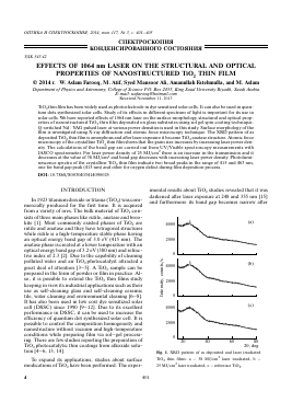
Fig. 1. XRD pattern of as deposited and laser irradiated TiO2 thin films: a — 38 MJ/cm2 laser irradiated, b — 25 MJ/cm2 laser irradiated, c — reference TiO2.
4
401
0
402
ASLAM FAROOQ et al.
nm 83.3.
nm 89.3.
As grown 25 MW/cm2 laser irradiated 38 MW/cm2 laser irradiated
Crystalline size from 23 28
XRD, nm
Grain size from 20 - 31
AFM, nm
Roughness, nm 23.5 - 21
Transmission (%) 71 75 69
at 400 nm
Transmission (%) 87 96 89
at 800 nm
Band gap,eV 3.3 3.28 3.26
FWHM - 0.58 0.29
1:Height
2.0 mm -85.5 nm
2.0 mm -84.2 nm
Fig. 2. Three and two dimensional AFM image of as deposited TiO2 thin film.
plasma treatment [16]. The main cause of these phenomena is due to significant decrease of oxygen in the TiO2 lattice formed [15, 16]. The studies regarding la-
Structural and optical results of as grown and laser irradiated TiO2 thin film
Fig. 3. Three and two dimensional AFM image of as deposited TiO2 thin film with 60 mJ laser irradiated.
ser irradiation experiments [15] show that one-photon absorption can cause the dissociation of Ti—O bonds to produce oxygen deficiencies. However surface modications can be analyzed from precision materials processing and fine patterning as the laser can be focused onto a small area of the material. The use of continuous wave (CW), long pulsed, or nanosecond lasers lead to heating affect and morphological changes only in the laser spot [17—23] due to which it's processing capability is limited.
In the present investigation, TiO2 thin films are synthesized on glass substrate using sol-gel spin coating technique and then the samples are exposed to Nd : YAG pulsed laser with 1064 nm wavelength using different power densities. We have studied TiO2 thin film keeping in view of its effect on the structure and optical properties. The microstructure, surface morphology and phase purity of samples have been experimentally characterized using X-ray diffractometer (XRD), atomic force microscope while the optical properties are analysed with spectrophotometer and spectrofluo-rometer.
0
Wavelength, nm
Fig. 4. Transmission spectra of as deposited and laser irradiated TiO2 thin films: 1 — 25 MJ/cm2 laser irradiated, 2 — 38 MJ/cm2 laser irradiated, 3 — reference film.
EXPERIMENTAL DETAILS
Titanium dioxide thin films were deposited on glass substrates by the spin coating method. Titanium (IV) oxide, a mixture of rutile and anatase nanoparticles with <250 nm particle size (DLS) paste, 53-57 wt. % in diethylene glycol monobutyl ether/ethylene glycol, 99.9% trace metals basis (700355) was purchased from Sigma Aldrich. The paste was further diluted with diethylene glycol and ethanol. The resulting paste was magnetic stirred at room temperature for 30 min followed by ultrasonification for fine mixing and the sol is ready for thin film deposition. Prior to deposition the glass substrates were thoroughly cleaned using standard cleaning methods. Glass substrates were soaked in chromic acid for overnight which then cleaned with detergent water and distilled water many times followed by acetone and ultrasonic cleaner. Finally the substrates were rinsed with distilled water and finally dried with pure nitrogen. TiO2 thin films were deposited on glass substrates using spincoat G3P-8 spincoater at 3250 rpm for 30 s and annealed at 350°C in air for 1 h. After synthesis, the TiO2 thin films samples are exposed to fundamental beam of Quantel Bril-liant/BrilliantB Q-switched Nd : YAG pulsed laser (Quantel-Brilliant) at 1064 nm with 8 ns pulse width and 7 mm diameter. The samples were irradiated at two different power densities 25 and 38 MW/cm2.
The microstructure of reference and laser irradiated TiO2 thin film samples was characterized by X-ray diffraction using multipurpose X-ray diffractometer (Bruker, D8 Advance) with Cu^ source radiation. The optical transmission of the samples was recorded at room temperature by a JASCO UV/VIS/NIR spectrophotometer (V-670) in the wavelength range of 280—850 nm. Photoluminescence measurement was recorded using JASCO spectrofluorometer (FP-8200). Surface morphology of the thin films was investigated with AFM (Quesant Universal SPM, Ambios Technology) in contact mode. An AFM tip of silicon nitride was used having an approximate radius of curvature of 10 nm.
RESULTS AND DISCUSSION
The XRD pattern of as grown and laser irradiated TiO2 thin film at different power densities deposited on the glass substrate are shown in Fig. 1. The XRD of the glass substrate is subtracted from the data. As grown TiO2 thin film is amorphous in nature and after laser exposure the peak (101) appeared at 26° which shows anatase phase of TiO2 thin film. The full width half maximum (FWHM) decreased and the crystallite increased by increasing the laser power density (Table).
The surface morphology of the as grown and laser irradiated TiO2 thin film were observed by atomic force
404
ASLAM FAROOQ et al.
0.02 r
0.01 -
0 0.02
0 0.02
0.01
hv, eV
Fig. 5. Tauc plot of as deposited (a) and laser irradiated TiO2 thin films: b - 25 MW/cm2, c - 38 MW/cm2.
microscope (AFM). Three and two dimensional AFM image of deposited TiO2 thin film are shown in Figs. 2 and 3. Root mean square (RMS) roughness of the films was extracted from AFM data. The RMS roughness of as grown TiO2 thin film was 23.5 nm and after the laser irradiation it is decreased to 21 nm. The experimental results of grain size are in good agreement with the XRD analysis revealing that the particles size is increased with increasing laser power density.
The optical transmittance of as grown and laser exposed TiO2 thin film was measured using UV-Visible spectroscopy and their spectra are depicted in Fig. 4. We found that the transmission in the visible range for as grown and laser irradiated thin film samples were
87-96%. A pure TiO2 thin film had low transmission and at 25 MW/cm2 the transmission increases significantly and reaches to 96%. If we increase the laser power density to 38 MW/cm2 there is a sharp decrease in the transmission of TiO2 thin film. This may be attributed to the surface roughness of the laser exposed samples confirmed by the AFM results.
To calculate band gap transition, the absorption coeffici
Для дальнейшего прочтения статьи необходимо приобрести полный текст. Статьи высылаются в формате PDF на указанную при оплате почту. Время доставки составляет менее 10 минут. Стоимость одной статьи — 150 рублей.